{"title":"Comparison of thoracic vertebrae and L1 CT attenuation in predicting osteoporosis using opportunistic chest CT.","authors":"Lilan Wu, Shunfa Huang, Liling Xu, Shengxiang Rao, Zhen Qian, Mengze Zhang, Ying Yuan, Jianjun Zhou","doi":"10.1177/1759720X251374134","DOIUrl":null,"url":null,"abstract":"<p><strong>Background: </strong>Since dual-energy x-ray absorptiometry (DXA) is currently the most commonly used reference standard, most previous studies using computed tomography (CT) attenuation values to predict osteoporosis have chosen abdominal CT images. A few studies have investigated whether the thoracic vertebrae can be independently used for the identification of osteoporosis compared to the lumbar vertebrae.</p><p><strong>Objective: </strong>To investigate whether the attenuation values of thoracic vertebrae measured using artificial intelligence (AI) on chest CT would independently predict osteoporosis identification, considering central DXA as a reference standard.</p><p><strong>Design: </strong>This was a cross-sectional study.</p><p><strong>Methods: </strong>A total of 553 participants (353 men and 200 women) who underwent chest CT and DXA within 1 day were included. The attenuation values (HU) of the T7-12 vertebrae and L1 vertebra were obtained by AI. The effects of the clinical baseline data and attenuation values among the normal, osteopenia, and osteoporosis groups were compared. The correlation between attenuation and bone mineral density (BMD) values was analyzed, and the diagnostic performance of thoracic and first lumbar vertebrae attenuation values for diagnosing osteopenia or osteoporosis was further explored.</p><p><strong>Results: </strong>The CT attenuation values of T7-12 and L1 vertebrae showed positive correlation with <i>T</i>-score (<i>R</i> = 0.58-0.61, <i>p</i> < 0.01). T12 attenuation >184.8 HU was 84.1% sensitive and 70.6% specific for distinguishing normal BMD, while T12 attenuation <146.2 HU was 61.4% specific and 75.6% sensitive for distinguishing osteoporosis from osteopenia. There were no significant differences between the T10-12 and L1 groups in distinguishing the normal, osteopenia, and osteoporosis groups. Moreover, the diagnostic efficacy among the T10, T11, T12, and L1 vertebral bodies was not statistically significantly different among the three groups.</p><p><strong>Conclusion: </strong>Opportunistic screening is a valid method for predicting osteopenia or osteoporosis. As a rapid and effective tool, T10-12 vertebral attenuation measures can be incorporated to predict osteoporosis and identify patients who may benefit from further investigations using DXA based on routine clinical chest CT examinations.</p>","PeriodicalId":23056,"journal":{"name":"Therapeutic Advances in Musculoskeletal Disease","volume":"17 ","pages":"1759720X251374134"},"PeriodicalIF":4.1000,"publicationDate":"2025-09-11","publicationTypes":"Journal Article","fieldsOfStudy":null,"isOpenAccess":false,"openAccessPdf":"https://www.ncbi.nlm.nih.gov/pmc/articles/PMC12432313/pdf/","citationCount":"0","resultStr":null,"platform":"Semanticscholar","paperid":null,"PeriodicalName":"Therapeutic Advances in Musculoskeletal Disease","FirstCategoryId":"3","ListUrlMain":"https://doi.org/10.1177/1759720X251374134","RegionNum":2,"RegionCategory":"医学","ArticlePicture":[],"TitleCN":null,"AbstractTextCN":null,"PMCID":null,"EPubDate":"2025/1/1 0:00:00","PubModel":"eCollection","JCR":"Q2","JCRName":"RHEUMATOLOGY","Score":null,"Total":0}
引用次数: 0
Abstract
Background: Since dual-energy x-ray absorptiometry (DXA) is currently the most commonly used reference standard, most previous studies using computed tomography (CT) attenuation values to predict osteoporosis have chosen abdominal CT images. A few studies have investigated whether the thoracic vertebrae can be independently used for the identification of osteoporosis compared to the lumbar vertebrae.
Objective: To investigate whether the attenuation values of thoracic vertebrae measured using artificial intelligence (AI) on chest CT would independently predict osteoporosis identification, considering central DXA as a reference standard.
Design: This was a cross-sectional study.
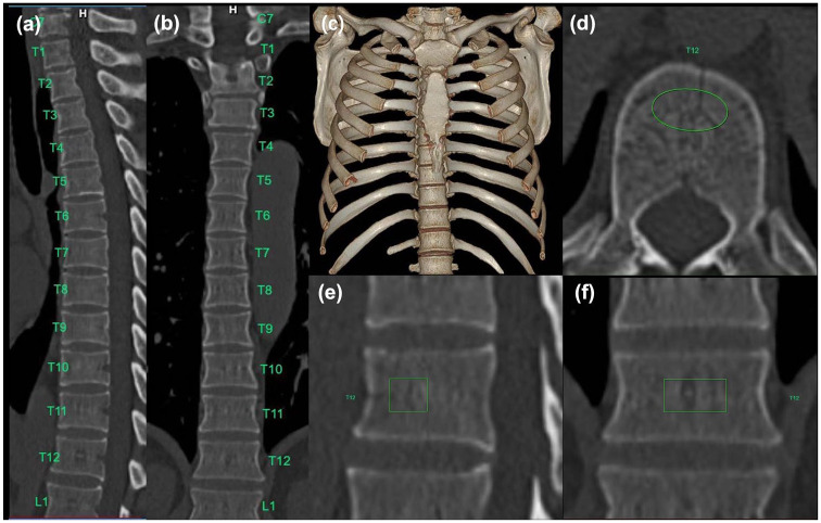
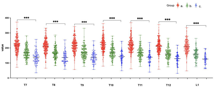
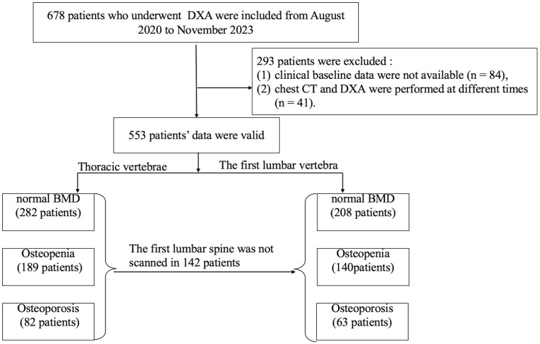
Methods: A total of 553 participants (353 men and 200 women) who underwent chest CT and DXA within 1 day were included. The attenuation values (HU) of the T7-12 vertebrae and L1 vertebra were obtained by AI. The effects of the clinical baseline data and attenuation values among the normal, osteopenia, and osteoporosis groups were compared. The correlation between attenuation and bone mineral density (BMD) values was analyzed, and the diagnostic performance of thoracic and first lumbar vertebrae attenuation values for diagnosing osteopenia or osteoporosis was further explored.
Results: The CT attenuation values of T7-12 and L1 vertebrae showed positive correlation with T-score (R = 0.58-0.61, p < 0.01). T12 attenuation >184.8 HU was 84.1% sensitive and 70.6% specific for distinguishing normal BMD, while T12 attenuation <146.2 HU was 61.4% specific and 75.6% sensitive for distinguishing osteoporosis from osteopenia. There were no significant differences between the T10-12 and L1 groups in distinguishing the normal, osteopenia, and osteoporosis groups. Moreover, the diagnostic efficacy among the T10, T11, T12, and L1 vertebral bodies was not statistically significantly different among the three groups.
Conclusion: Opportunistic screening is a valid method for predicting osteopenia or osteoporosis. As a rapid and effective tool, T10-12 vertebral attenuation measures can be incorporated to predict osteoporosis and identify patients who may benefit from further investigations using DXA based on routine clinical chest CT examinations.
期刊介绍:
Therapeutic Advances in Musculoskeletal Disease delivers the highest quality peer-reviewed articles, reviews, and scholarly comment on pioneering efforts and innovative studies across all areas of musculoskeletal disease.
 求助内容:
求助内容: 应助结果提醒方式:


