Multimodal model enhances qualitative diagnosis of hypervascular thyroid nodules: integrating radiomics and deep learning features based on B-mode and PDI images.
{"title":"Multimodal model enhances qualitative diagnosis of hypervascular thyroid nodules: integrating radiomics and deep learning features based on B-mode and PDI images.","authors":"Wen Wen, Tingrui Zhang, Haina Zhao, Jingyan Liu, Heng Jiang, Yushuang He, Zekun Jiang","doi":"10.21037/gs-2025-183","DOIUrl":null,"url":null,"abstract":"<p><strong>Background: </strong>Facing challenges in differentiating benign/malignant hypervascular thyroid nodules due to overlapping ultrasound features and limited vascular characterization, this study developed multimodal machine learning models integrating B-mode and power Doppler imaging (PDI) features.</p><p><strong>Methods: </strong>A retrospective cohort of 315 patients with pathologically confirmed hypervascular thyroid nodules (Adler grade 2/3) was divided into training (n=220) and test (n=95) sets. Multimodal ultrasound images were processed using a deep learning-based segmentation model and red-channel thresholding method, followed by radiomics feature extraction (1,910 features via PyRadiomics) and deep learning feature derivation (1,000 ResNet-derived features). Feature selection employed analysis of variance (ANOVA) F-tests, yielding hybrid feature sets. Five machine learning algorithms, including random forest, logistic regression, support vector machine (SVM), eXtreme Gradient Boosting (XGBoost), and Tabular Prior-data Fitted Network (TABPFN), were trained and validated. A fused model integrated optimal B-mode and PDI SVM models. Performance was assessed via area under the curve (AUC), accuracy, precision, recall, and SHapley Additive exPlanations (SHAP) analysis. Clinical trial registration number: ChinCTR2100049742.</p><p><strong>Results: </strong>SVM outperformed other models in single-modality analyses: B-mode SVM achieved an AUC of 0.89 (accuracy: 0.84; recall: 0.94), while PDI SVM attained an AUC of 0.86 (accuracy: 0.82; recall: 0.97). The combined model demonstrated near-perfect training performance (AUC: 1.00; accuracy: 0.96) but moderated in testing (AUC: 0.89; accuracy: 0.78), indicating potential overfitting. Radiomics features dominated feature importance, including Logarithm_firstorder_Energy (B-mode) and squareroot_firstorder_Minimum (PDI). The fused model showed superior recall (0.95) and F1-score (0.86) compared to single modalities, highlighting complementary diagnostic value.</p><p><strong>Conclusions: </strong>Multimodal ultrasound fusion models, particularly SVM-based frameworks, enhance diagnostic accuracy for hypervascular thyroid nodules by synergizing morphological and vascular features. Despite challenges in generalizability, the integration of radiomics and deep learning features offers clinically reliable tools to reduce invasive biopsies.</p>","PeriodicalId":12760,"journal":{"name":"Gland surgery","volume":"14 8","pages":"1558-1571"},"PeriodicalIF":1.6000,"publicationDate":"2025-08-31","publicationTypes":"Journal Article","fieldsOfStudy":null,"isOpenAccess":false,"openAccessPdf":"https://www.ncbi.nlm.nih.gov/pmc/articles/PMC12432950/pdf/","citationCount":"0","resultStr":null,"platform":"Semanticscholar","paperid":null,"PeriodicalName":"Gland surgery","FirstCategoryId":"3","ListUrlMain":"https://doi.org/10.21037/gs-2025-183","RegionNum":3,"RegionCategory":"医学","ArticlePicture":[],"TitleCN":null,"AbstractTextCN":null,"PMCID":null,"EPubDate":"2025/8/26 0:00:00","PubModel":"Epub","JCR":"Q3","JCRName":"SURGERY","Score":null,"Total":0}
引用次数: 0
Abstract
Background: Facing challenges in differentiating benign/malignant hypervascular thyroid nodules due to overlapping ultrasound features and limited vascular characterization, this study developed multimodal machine learning models integrating B-mode and power Doppler imaging (PDI) features.
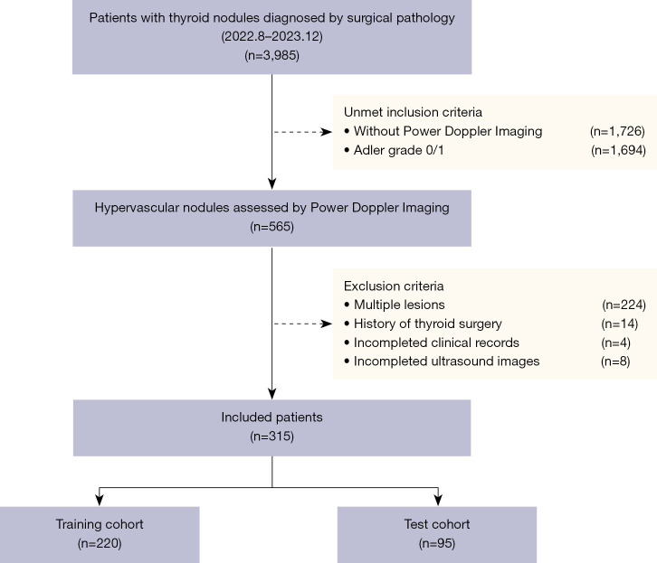
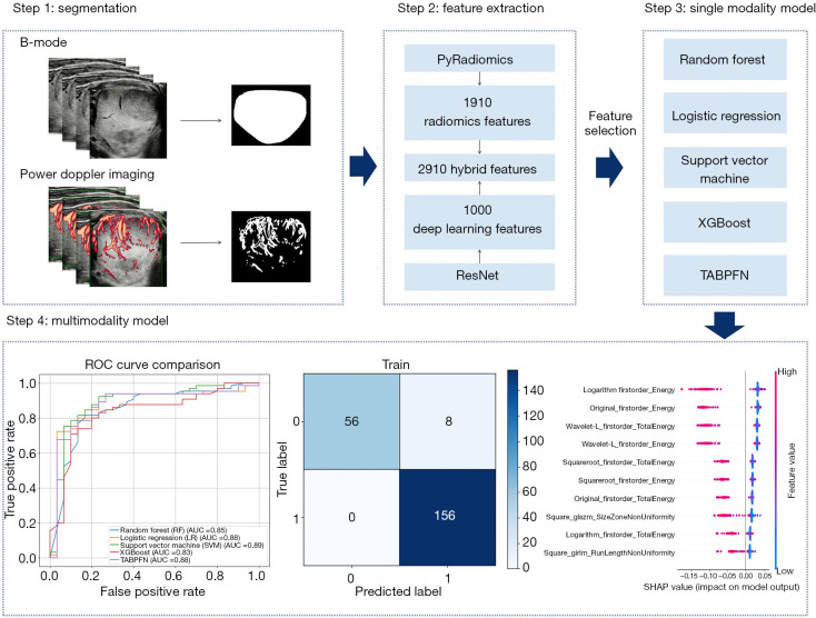
Methods: A retrospective cohort of 315 patients with pathologically confirmed hypervascular thyroid nodules (Adler grade 2/3) was divided into training (n=220) and test (n=95) sets. Multimodal ultrasound images were processed using a deep learning-based segmentation model and red-channel thresholding method, followed by radiomics feature extraction (1,910 features via PyRadiomics) and deep learning feature derivation (1,000 ResNet-derived features). Feature selection employed analysis of variance (ANOVA) F-tests, yielding hybrid feature sets. Five machine learning algorithms, including random forest, logistic regression, support vector machine (SVM), eXtreme Gradient Boosting (XGBoost), and Tabular Prior-data Fitted Network (TABPFN), were trained and validated. A fused model integrated optimal B-mode and PDI SVM models. Performance was assessed via area under the curve (AUC), accuracy, precision, recall, and SHapley Additive exPlanations (SHAP) analysis. Clinical trial registration number: ChinCTR2100049742.
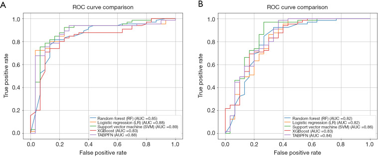
Results: SVM outperformed other models in single-modality analyses: B-mode SVM achieved an AUC of 0.89 (accuracy: 0.84; recall: 0.94), while PDI SVM attained an AUC of 0.86 (accuracy: 0.82; recall: 0.97). The combined model demonstrated near-perfect training performance (AUC: 1.00; accuracy: 0.96) but moderated in testing (AUC: 0.89; accuracy: 0.78), indicating potential overfitting. Radiomics features dominated feature importance, including Logarithm_firstorder_Energy (B-mode) and squareroot_firstorder_Minimum (PDI). The fused model showed superior recall (0.95) and F1-score (0.86) compared to single modalities, highlighting complementary diagnostic value.
Conclusions: Multimodal ultrasound fusion models, particularly SVM-based frameworks, enhance diagnostic accuracy for hypervascular thyroid nodules by synergizing morphological and vascular features. Despite challenges in generalizability, the integration of radiomics and deep learning features offers clinically reliable tools to reduce invasive biopsies.
期刊介绍:
Gland Surgery (Gland Surg; GS, Print ISSN 2227-684X; Online ISSN 2227-8575) being indexed by PubMed/PubMed Central, is an open access, peer-review journal launched at May of 2012, published bio-monthly since February 2015.
 求助内容:
求助内容: 应助结果提醒方式:


