Improved diagnostic value of whole-lesion histogram and texture analyses on multiparametric breast MRI for papillary neoplasms with non-mass enhancement.
Xinyue Li, Qiuyi Fu, Kun Sun, Fuhua Yan, Weimin Chai
{"title":"Improved diagnostic value of whole-lesion histogram and texture analyses on multiparametric breast MRI for papillary neoplasms with non-mass enhancement.","authors":"Xinyue Li, Qiuyi Fu, Kun Sun, Fuhua Yan, Weimin Chai","doi":"10.21037/gs-2025-128","DOIUrl":null,"url":null,"abstract":"<p><strong>Background: </strong>Differentiating between benign and malignant entities remains a complex aspect in the diagnosis of breast papillary neoplasms. This study aimed to assess if analyzing whole-lesion histograms and texture features on multiparametric magnetic resonance imaging (MRI) can enhance the diagnostic accuracy of breast papillary neoplasms presenting as non-mass enhancement (NME).</p><p><strong>Methods: </strong>In this retrospective analysis, 98 female patients with 98 papillary neoplasms exhibiting NME on dynamic contrast-enhanced (DCE) MRI were enrolled. Two radiologists independently assessed all lesions and later established a consensus on morphological features based on the Breast Imaging Reporting and Data System (BI-RADS) criteria. Quantitative histogram and texture metrics were extracted from four MRI sequences: diffusion-weighted imaging (DWI) with b values of 50 and 1,000 s/mm<sup>2</sup>, apparent diffusion coefficient (ADC) map, and contrast-enhanced T1-weighted subtraction (SUB) magnetic resonance (MR) images. The least absolute shrinkage and selection operator (LASSO) was applied to feature selection. A multivariable logistic regression model was developed using stepwise covariate selection. Diagnostic efficacy was assessed via receiver operating characteristic (ROC) curve analysis.</p><p><strong>Results: </strong>According to BI-RADS, benign and malignant papillary neoplasms with NME differed significantly in the amount of fibroglandular tissue (FGT), distribution, and time-intensity curve (TIC) pattern (P=0.04, 0.008, <0.001, respectively), yielding an area under the ROC curve (AUC) of 0.792 (sensitivity 67.4%, specificity 84.6%). Quantitative analysis revealed differences in the ADC<sub>standard deviation (SD)</sub>, ADC<sub>5th percentile</sub>, ADC<sub>differential entropy (diff-entropy)</sub>, ADC<sub>contrast</sub>, DWI<sub>b50-SD</sub>, DWI<sub>b800-mean</sub>, and SUB MR<sub>95th percentile</sub> (P=0.009, 0.01, 0.001, 0.01, 0.001, 0.002, 0.02, respectively), achieving an AUC of 0.908 (sensitivity 82.6%, specificity 88.5%). The AUC of the quantitative model outperformed that of the qualitative model (P<0.001). The AUC of the quantitative model for distinguishing malignant NME papillary neoplasms from benign NME papillary neoplasms in the internal validation set was 0.941, with a sensitivity of 90.4%, and a specificity of 87.0%.</p><p><strong>Conclusions: </strong>Compared to the qualitative BI-RADS assessment, quantitative analysis of whole-lesion histogram and texture on multiparametric MRI is proven to be more effective in distinguishing between benign and malignant papillary breast neoplasms with NME, in order to avoid overtreatment.</p>","PeriodicalId":12760,"journal":{"name":"Gland surgery","volume":"14 8","pages":"1444-1455"},"PeriodicalIF":1.6000,"publicationDate":"2025-08-31","publicationTypes":"Journal Article","fieldsOfStudy":null,"isOpenAccess":false,"openAccessPdf":"https://www.ncbi.nlm.nih.gov/pmc/articles/PMC12432942/pdf/","citationCount":"0","resultStr":null,"platform":"Semanticscholar","paperid":null,"PeriodicalName":"Gland surgery","FirstCategoryId":"3","ListUrlMain":"https://doi.org/10.21037/gs-2025-128","RegionNum":3,"RegionCategory":"医学","ArticlePicture":[],"TitleCN":null,"AbstractTextCN":null,"PMCID":null,"EPubDate":"2025/8/18 0:00:00","PubModel":"Epub","JCR":"Q3","JCRName":"SURGERY","Score":null,"Total":0}
引用次数: 0
Abstract
Background: Differentiating between benign and malignant entities remains a complex aspect in the diagnosis of breast papillary neoplasms. This study aimed to assess if analyzing whole-lesion histograms and texture features on multiparametric magnetic resonance imaging (MRI) can enhance the diagnostic accuracy of breast papillary neoplasms presenting as non-mass enhancement (NME).
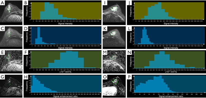
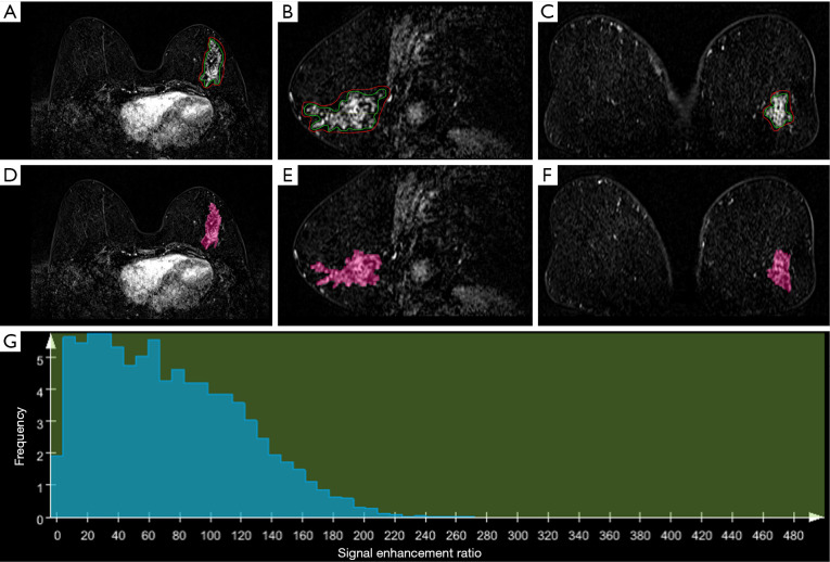
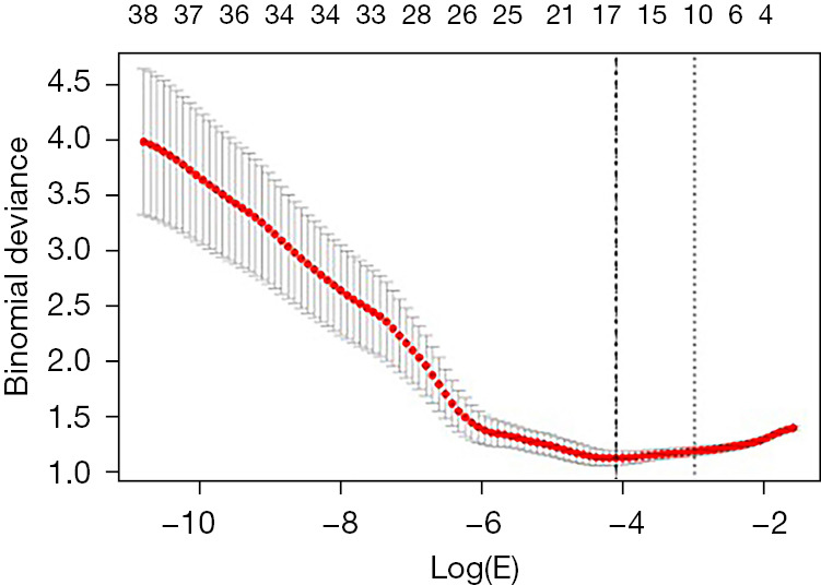
Methods: In this retrospective analysis, 98 female patients with 98 papillary neoplasms exhibiting NME on dynamic contrast-enhanced (DCE) MRI were enrolled. Two radiologists independently assessed all lesions and later established a consensus on morphological features based on the Breast Imaging Reporting and Data System (BI-RADS) criteria. Quantitative histogram and texture metrics were extracted from four MRI sequences: diffusion-weighted imaging (DWI) with b values of 50 and 1,000 s/mm2, apparent diffusion coefficient (ADC) map, and contrast-enhanced T1-weighted subtraction (SUB) magnetic resonance (MR) images. The least absolute shrinkage and selection operator (LASSO) was applied to feature selection. A multivariable logistic regression model was developed using stepwise covariate selection. Diagnostic efficacy was assessed via receiver operating characteristic (ROC) curve analysis.
Results: According to BI-RADS, benign and malignant papillary neoplasms with NME differed significantly in the amount of fibroglandular tissue (FGT), distribution, and time-intensity curve (TIC) pattern (P=0.04, 0.008, <0.001, respectively), yielding an area under the ROC curve (AUC) of 0.792 (sensitivity 67.4%, specificity 84.6%). Quantitative analysis revealed differences in the ADCstandard deviation (SD), ADC5th percentile, ADCdifferential entropy (diff-entropy), ADCcontrast, DWIb50-SD, DWIb800-mean, and SUB MR95th percentile (P=0.009, 0.01, 0.001, 0.01, 0.001, 0.002, 0.02, respectively), achieving an AUC of 0.908 (sensitivity 82.6%, specificity 88.5%). The AUC of the quantitative model outperformed that of the qualitative model (P<0.001). The AUC of the quantitative model for distinguishing malignant NME papillary neoplasms from benign NME papillary neoplasms in the internal validation set was 0.941, with a sensitivity of 90.4%, and a specificity of 87.0%.
Conclusions: Compared to the qualitative BI-RADS assessment, quantitative analysis of whole-lesion histogram and texture on multiparametric MRI is proven to be more effective in distinguishing between benign and malignant papillary breast neoplasms with NME, in order to avoid overtreatment.
期刊介绍:
Gland Surgery (Gland Surg; GS, Print ISSN 2227-684X; Online ISSN 2227-8575) being indexed by PubMed/PubMed Central, is an open access, peer-review journal launched at May of 2012, published bio-monthly since February 2015.
 求助内容:
求助内容: 应助结果提醒方式:


