Automated quantification of collagen proportionate area correlates with molecular and histological markers of fibrosis in CCl4-treated rats
IF 3.7
4区 医学
Q2 PATHOLOGY
引用次数: 0
Abstract
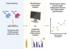
Liver fibrosis results from chronic liver injury and is characterized by excessive accumulation of extracellular matrix due to sustained wound-healing responses. Although histological evaluation remains the gold standard for fibrosis assessment, its subjectivity can limit reproducibility. In this study, we evaluated an automated image analysis software, MorphoQuant, for liver fibrosis quantification in a rat model of CCl4-induced liver injury. Male Wistar rats were treated with CCl4 or vehicle for six weeks, and fibrosis severity was assessed using both the conventional Ishak staging system and automated quantification of collagen proportionate area (CPA). Automated CPA strongly correlated with Ishak stage, liver index, and plasma aminotransferase levels. Additionally, CPA values were significantly associated with the expression of fibrosis-related genes and macrophage infiltration, highlighting the software's ability to assess both fibrosis progression and inflammatory responses. These findings support the use of MorphoQuant as a robust, reader-independent tool that enhance analytical consistency in preclinical models of liver fibrosis.
胶原比例面积的自动定量与ccl4治疗大鼠纤维化的分子和组织学标志物相关
肝纤维化是由慢性肝损伤引起的,其特征是由于持续的伤口愈合反应导致细胞外基质过度积累。尽管组织学评估仍然是纤维化评估的金标准,但其主观性限制了可重复性。在这项研究中,我们评估了自动图像分析软件MorphoQuant在ccl4诱导的肝损伤大鼠模型中的肝纤维化定量。雄性Wistar大鼠用CCl4或载药治疗6周,使用传统的Ishak分期系统和胶原比例面积(CPA)自动定量评估纤维化严重程度。自动CPA与Ishak分期、肝脏指数和血浆转氨酶水平密切相关。此外,CPA值与纤维化相关基因的表达和巨噬细胞浸润显著相关,突出了该软件评估纤维化进展和炎症反应的能力。这些发现支持MorphoQuant作为一种强大的、独立于阅读器的工具,可以增强肝纤维化临床前模型的分析一致性。
本文章由计算机程序翻译,如有差异,请以英文原文为准。
求助全文
约1分钟内获得全文
求助全文
来源期刊
CiteScore
8.90
自引率
0.00%
发文量
78
审稿时长
11.5 weeks
期刊介绍:
Under new editorial leadership, Experimental and Molecular Pathology presents original articles on disease processes in relation to structural and biochemical alterations in mammalian tissues and fluids and on the application of newer techniques of molecular biology to problems of pathology in humans and other animals. The journal also publishes selected interpretive synthesis reviews by bench level investigators working at the "cutting edge" of contemporary research in pathology. In addition, special thematic issues present original research reports that unravel some of Nature''s most jealously guarded secrets on the pathologic basis of disease.
Research Areas include: Stem cells; Neoangiogenesis; Molecular diagnostics; Polymerase chain reaction; In situ hybridization; DNA sequencing; Cell receptors; Carcinogenesis; Pathobiology of neoplasia; Complex infectious diseases; Transplantation; Cytokines; Flow cytomeric analysis; Inflammation; Cellular injury; Immunology and hypersensitivity; Athersclerosis.
 求助内容:
求助内容: 应助结果提醒方式:


