Spatio-temporal registration of multi-perspective 3D echocardiography for improved strain estimation
IF 11.8
1区 医学
Q1 COMPUTER SCIENCE, ARTIFICIAL INTELLIGENCE
引用次数: 0
Abstract
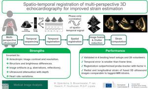
For heart diagnostics, ultrasound is generally the modality of choice due to its high temporal and spatial resolution, availability, and patient safety. Although 3D echocardiography captures the complex shape and motion of the heart with more precision than 2D, it suffers to a greater extent from poor resolution, noise, and limited field-of-view. Multi-perspective echocardiography has proven to significantly enhance both image quality and field-of-view. The greatest improvements occur when combining acquisitions from widely differing insonification angles, but this process is challenging because of substantial local structural and brightness variations and ultrasound’s anisotropic nature. To handle these inconsistencies, a novel temporal and spatial registration algorithm designed is proposed. Temporal registration is achieved using low-frequency cardiac wall features and motion extracted via singular value decomposition of a spatio-temporal Casorati matrix, while spatial registration is performed using phase-only correlation of low-frequency data. The acquisitions are seamlessly fused using a 3D, oriented, wavelet transform including a near-field clutter algorithm. In vitro and in vivo testing highlights the benefits of this approach. Temporal alignment, validated against electrocardiograms, is precise, with an average error of just 2 ± 10 ms. Furthermore, our method outperforms a six-degree-of-freedom encoder-based probe tracker, reducing spatial registration error to 5 ± 3 mm from 19 ± 10 mm. The resulting longitudinal and radial strain measurements closely align with those obtained by tagged magnetic resonance imaging, demonstrating the accuracy and feasibility of this technique.
多视角三维超声心动图的时空配准改进应变估计
对于心脏诊断,超声通常是首选的方式,因为它具有高时间和空间分辨率,可用性和患者安全性。尽管3D超声心动图比2D更精确地捕捉心脏的复杂形状和运动,但它在更大程度上受到分辨率差、噪音和视野有限的影响。多视角超声心动图已被证明可以显著提高图像质量和视野。最大的改进发生在从广泛不同的失谐角度合并采集时,但由于大量的局部结构和亮度变化以及超声的各向异性,这一过程具有挑战性。为了处理这些不一致,设计了一种新的时空配准算法。通过时空Casorati矩阵的奇异值分解提取低频心壁特征和运动来实现时间配准,而空间配准则使用低频数据的纯相位相关进行。利用三维定向小波变换(包括近场杂波算法)无缝融合采集的数据。体外和体内测试突出了这种方法的好处。根据心电图验证的时间对齐是精确的,平均误差仅为2±10毫秒。此外,我们的方法优于基于六自由度编码器的探针跟踪器,将空间配准误差从19±10 mm减少到5±3 mm。由此产生的纵向和径向应变测量结果与标记磁共振成像获得的结果密切一致,证明了该技术的准确性和可行性。
本文章由计算机程序翻译,如有差异,请以英文原文为准。
求助全文
约1分钟内获得全文
求助全文
来源期刊

Medical image analysis
工程技术-工程:生物医学
CiteScore
22.10
自引率
6.40%
发文量
309
审稿时长
6.6 months
期刊介绍:
Medical Image Analysis serves as a platform for sharing new research findings in the realm of medical and biological image analysis, with a focus on applications of computer vision, virtual reality, and robotics to biomedical imaging challenges. The journal prioritizes the publication of high-quality, original papers contributing to the fundamental science of processing, analyzing, and utilizing medical and biological images. It welcomes approaches utilizing biomedical image datasets across all spatial scales, from molecular/cellular imaging to tissue/organ imaging.
 求助内容:
求助内容: 应助结果提醒方式:


