Metal artifact reduction from surgical clips for intracranial aneurysms in photon-counting detector CT angiography
IF 3.3
3区 医学
Q1 RADIOLOGY, NUCLEAR MEDICINE & MEDICAL IMAGING
引用次数: 0
Abstract
Objectives
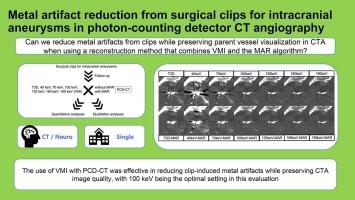
To identify conditions that reduce metal artifacts from surgical clips for intracranial aneurysms while preserving parent vessel visualization in CT angiography (CTA) by using a reconstruction method combining virtual monoenergetic image (VMI) and a metal artifact reduction (MAR) algorithm.
Materials & Methods
Patients who underwent CTA after clipping surgery using photon-counting detector CT (PCD-CT) in 2023–2025 were included. Images were reconstructed as conventional polychromatic 120kVp images (T3D) and 40, 70, 100, 130, 160, and 190 keV using VMI; we created images applying the MAR algorithm to each. Hyperdense/ hypodense artifacts, the artifact index (AI), and the contrast-to-noise ratio (CNR) were quantitatively measured by two neuroradiologists. Hyperdense/ hypodense artifacts, noise, parent vessel visualization, blooming, and new artifacts generated with the MAR algorithm were qualitatively assessed by four neuroradiologists.
Results
28 patients (median 64 [53–72] years, 15 men) were included. Quantitatively, hyperdense artifacts decreased at ≥ 70 keV (68.4H.U.) versus T3D (82.7) (P < 0.001). Hypodense artifacts and AI decreased at ≥ 100 keV (-2.3H.U. and 21.6) (P < 0.001). CNRs decreased at ≥ 70 keV (20.1) and further decreased when the MAR was applied (13.6) (P < 0.001). Qualitatively, hyperdense/ hypodense artifacts decreased at ≥ 100 keV and further decreased slightly with the MAR (P < 0.001). Noise decreased at ≥ 100 keV. The parent vessel visualization after clipping improved at 70 and 100 keV but decreased with the MAR (P < 0.001). Clip blooming did not occur at ≥ 100 keV (P < 0.001) regardless of the use/nonuse of the MAR (P > 0.500). New artifacts decreased at ≥ 100 keV-MAR versus T3D-MAR (P < 0.001).
Conclusion
The optimal setting for reducing metal artifacts while preserving vascular information was 100 keV.
光子计数检测器CT血管造影中颅内动脉瘤手术夹金属伪影的减少
目的采用虚拟单能成像(VMI)和金属伪影还原(MAR)算法相结合的重建方法,探讨在保留CT血管造影(CTA)显示的同时,减少颅内动脉瘤手术夹产生的金属伪影的条件。方法选取2023 ~ 2025年期间使用光子计数检测器CT (PCD-CT)进行夹持术后CTA的患者。利用VMI将图像重构为120kVp (T3D)和40,70,100,130,160和190kev的常规多色图像;我们将MAR算法应用于每个图像。由两名神经放射学家定量测量高密度/低密度伪影、伪影指数(AI)和噪声对比比(CNR)。由四名神经放射学家对使用MAR算法产生的高密度/低密度伪影、噪声、母血管可视化、虚化和新伪影进行定性评估。结果纳入28例患者,中位年龄64[53-72]岁,男性15例。在定量上,高密度伪影在≥70 keV (68.4H.U)时与T3D (82.7 h . u)相比减少(P < 0.001)。低密度伪影和AI在≥100 keV (-2.3H.U)时降低。21.6) (P < 0.001)。cnr在≥70 keV时下降(20.1),在施加MAR时进一步下降(13.6)(P < 0.001)。定性地说,高密度/低密度伪影在≥100 keV时减少,并随着MAR进一步略有减少(P < 0.001)。噪声在≥100 keV时降低。在70和100 keV时,夹持后的母血管可视化得到改善,但随着MAR的降低(P < 0.001)。无论是否使用MAR (P > 0.500),在≥100 keV时均未发生夹花(P < 0.001)。与T3D-MAR相比,≥100 kv - mar时新的伪影减少(P < 0.001)。结论在保留血管信息的同时减少金属伪影的最佳设置为100 keV。
本文章由计算机程序翻译,如有差异,请以英文原文为准。
求助全文
约1分钟内获得全文
求助全文
来源期刊

CiteScore
6.70
自引率
3.00%
发文量
398
审稿时长
42 days
期刊介绍:
European Journal of Radiology is an international journal which aims to communicate to its readers, state-of-the-art information on imaging developments in the form of high quality original research articles and timely reviews on current developments in the field.
Its audience includes clinicians at all levels of training including radiology trainees, newly qualified imaging specialists and the experienced radiologist. Its aim is to inform efficient, appropriate and evidence-based imaging practice to the benefit of patients worldwide.
 求助内容:
求助内容: 应助结果提醒方式:


