{"title":"Isolated Double Hoffa Fracture of the Distal Femur without Intercondylar Extension: A Rare Case Report.","authors":"Utkarsh Jain, Vipin Gupta, Navdeep Singh Keer","doi":"10.13107/jocr.2025.v15.i09.6018","DOIUrl":null,"url":null,"abstract":"<p><strong>Introduction: </strong>Hoffa fractures are rare intra-articular fractures of the distal femur in the coronal plane, typically affecting a single femoral condyle. A bicondylar involvement, essentially double Hoffa fractures, is exceedingly uncommon and rarely documented, especially in the absence of metaphyseal comminution.</p><p><strong>Case report: </strong>We present the case of a 25-year-old male who sustained an isolated bicondylar Hoffa fracture following a motorcycle accident. Radiographs and computed tomography (CT) imaging confirmed isolated coronal plane fractures of both femoral condyles (AO 33-B3), without metaphyseal comminution or intercondylar extension. The fracture was managed through a single medial parapatellar approach, using 5 cannulated screws and 2 headless Herbert screws. Post-operative rehabilitation involved early mobilization and progressive weight bearing. At 2 years follow-up, the patient demonstrated excellent functional recovery with a full range of motion (0-140°), no extensor lag, and pain-free full weight-bearing.</p><p><strong>Conclusion: </strong>This case highlights the role of precise anatomical reduction, CT evaluation, and early mobilization in managing complex distal femur fractures. This report adds to the limited literature on bicondylar Hoffa fractures without metaphyseal comminution.</p>","PeriodicalId":16647,"journal":{"name":"Journal of Orthopaedic Case Reports","volume":"15 9","pages":"66-70"},"PeriodicalIF":0.0000,"publicationDate":"2025-09-01","publicationTypes":"Journal Article","fieldsOfStudy":null,"isOpenAccess":false,"openAccessPdf":"https://www.ncbi.nlm.nih.gov/pmc/articles/PMC12422678/pdf/","citationCount":"0","resultStr":null,"platform":"Semanticscholar","paperid":null,"PeriodicalName":"Journal of Orthopaedic Case Reports","FirstCategoryId":"1085","ListUrlMain":"https://doi.org/10.13107/jocr.2025.v15.i09.6018","RegionNum":0,"RegionCategory":null,"ArticlePicture":[],"TitleCN":null,"AbstractTextCN":null,"PMCID":null,"EPubDate":"","PubModel":"","JCR":"","JCRName":"","Score":null,"Total":0}
引用次数: 0
Abstract
Introduction: Hoffa fractures are rare intra-articular fractures of the distal femur in the coronal plane, typically affecting a single femoral condyle. A bicondylar involvement, essentially double Hoffa fractures, is exceedingly uncommon and rarely documented, especially in the absence of metaphyseal comminution.
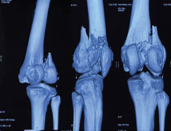
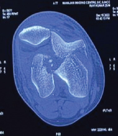
Case report: We present the case of a 25-year-old male who sustained an isolated bicondylar Hoffa fracture following a motorcycle accident. Radiographs and computed tomography (CT) imaging confirmed isolated coronal plane fractures of both femoral condyles (AO 33-B3), without metaphyseal comminution or intercondylar extension. The fracture was managed through a single medial parapatellar approach, using 5 cannulated screws and 2 headless Herbert screws. Post-operative rehabilitation involved early mobilization and progressive weight bearing. At 2 years follow-up, the patient demonstrated excellent functional recovery with a full range of motion (0-140°), no extensor lag, and pain-free full weight-bearing.
Conclusion: This case highlights the role of precise anatomical reduction, CT evaluation, and early mobilization in managing complex distal femur fractures. This report adds to the limited literature on bicondylar Hoffa fractures without metaphyseal comminution.


 求助内容:
求助内容: 应助结果提醒方式:


