S Venkatesh Kumar, Ashwath Ahila Baskar, K G Sathyendra, Ramson Vasagan, Rupali Dnyandeo Solankey, Rohini Venkatesh
{"title":"A Rare Case of Talus Neck and Open Medial Malleolus Fractures with Ankle Subluxation Treated using a Single Anteromedial Approach.","authors":"S Venkatesh Kumar, Ashwath Ahila Baskar, K G Sathyendra, Ramson Vasagan, Rupali Dnyandeo Solankey, Rohini Venkatesh","doi":"10.13107/jocr.2025.v15.i09.6098","DOIUrl":null,"url":null,"abstract":"<p><strong>Introduction: </strong>Talus fractures are uncommon and complex injuries associated with significant trauma and complications. The incidence of associated malleolar injury with talus fracture is rare.</p><p><strong>Case report: </strong>We share this unusual case of a Hawkins type-3 talus neck fracture along with a serious Grade 3B medial malleolus fracture and ankle subluxation, which was treated with cleaning the wound, realigning the ankle, and surgery to fix the bones. Post-operatively, the wound was healthy and free of infection. Despite being told to avoid weight-bearing for three months, the patient lost follow-up after a month and started occasional partial weight bearing. During the 10th post-operative week, we found a mild degree of talar neck collapse and Hawkins sign radiologically. The range of motion for the ankle was dorsiflexion of 0-15° and plantar flexion of 0-30°, with minimal swelling and pain on weight bearing.</p><p><strong>Conclusion: </strong>This case highlights the rarity and complexity of a talar neck fracture with ipsilateral medial malleolar fracture and ankle dislocation. Positive early outcomes were achieved through timely surgery within 10 h, careful soft tissue management, and appropriate fixation. The presence of a partial Hawkins sign post-operatively indicated preserved talar vascularity and reduced risk of avascular necrosis.</p>","PeriodicalId":16647,"journal":{"name":"Journal of Orthopaedic Case Reports","volume":"15 9","pages":"277-281"},"PeriodicalIF":0.0000,"publicationDate":"2025-09-01","publicationTypes":"Journal Article","fieldsOfStudy":null,"isOpenAccess":false,"openAccessPdf":"https://www.ncbi.nlm.nih.gov/pmc/articles/PMC12422633/pdf/","citationCount":"0","resultStr":null,"platform":"Semanticscholar","paperid":null,"PeriodicalName":"Journal of Orthopaedic Case Reports","FirstCategoryId":"1085","ListUrlMain":"https://doi.org/10.13107/jocr.2025.v15.i09.6098","RegionNum":0,"RegionCategory":null,"ArticlePicture":[],"TitleCN":null,"AbstractTextCN":null,"PMCID":null,"EPubDate":"","PubModel":"","JCR":"","JCRName":"","Score":null,"Total":0}
引用次数: 0
Abstract
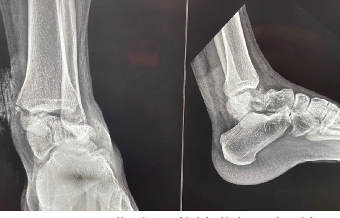
Introduction: Talus fractures are uncommon and complex injuries associated with significant trauma and complications. The incidence of associated malleolar injury with talus fracture is rare.
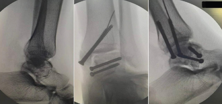
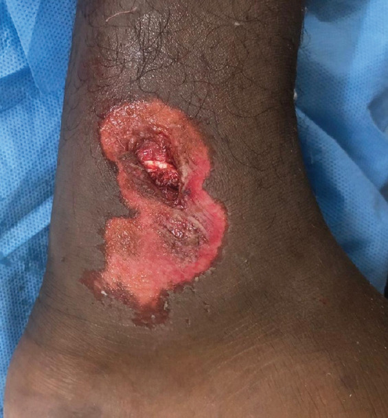
Case report: We share this unusual case of a Hawkins type-3 talus neck fracture along with a serious Grade 3B medial malleolus fracture and ankle subluxation, which was treated with cleaning the wound, realigning the ankle, and surgery to fix the bones. Post-operatively, the wound was healthy and free of infection. Despite being told to avoid weight-bearing for three months, the patient lost follow-up after a month and started occasional partial weight bearing. During the 10th post-operative week, we found a mild degree of talar neck collapse and Hawkins sign radiologically. The range of motion for the ankle was dorsiflexion of 0-15° and plantar flexion of 0-30°, with minimal swelling and pain on weight bearing.
Conclusion: This case highlights the rarity and complexity of a talar neck fracture with ipsilateral medial malleolar fracture and ankle dislocation. Positive early outcomes were achieved through timely surgery within 10 h, careful soft tissue management, and appropriate fixation. The presence of a partial Hawkins sign post-operatively indicated preserved talar vascularity and reduced risk of avascular necrosis.

 求助内容:
求助内容: 应助结果提醒方式:


