An Anomalous Attachment of the Medial Meniscus Posterior and Anterior Roots Mimicking a Bucket Handle Meniscus Tear: A Case Report and Review of the Literature.
Alexander J Egol, Bradley A Lezak, Emily Berzolla, Spencer M Stein
{"title":"An Anomalous Attachment of the Medial Meniscus Posterior and Anterior Roots Mimicking a Bucket Handle Meniscus Tear: A Case Report and Review of the Literature.","authors":"Alexander J Egol, Bradley A Lezak, Emily Berzolla, Spencer M Stein","doi":"10.13107/jocr.2025.v15.i09.6100","DOIUrl":null,"url":null,"abstract":"<p><strong>Introduction: </strong>Anomalous medial menisci are rare entities compared to their lateral counterparts. These anomalies include atypical insertions, most commonly into the anterior cruciate ligament, and discoid variants among others. This case adds to the literature on anomalous medial menisci with the presentation of a variant not described in the literature before.</p><p><strong>Case report: </strong>Our patient is a 38-year-old female who presented to the outpatient orthopedic clinic complaining of right knee pain in the setting of a traumatic kneeling event. She had a past medical history of asthma, polycystic ovary syndrome, and anxiety, but no surgical history related to the knee. She underwent magnetic resonance imaging identified what appeared to be a bucket-handle medial meniscus tear. The patient was brought to the operating room where diagnostic arthroscopy revealed a radial tear at the posterior horn of the medial meniscus as well as an anomalous connection between the anterior and posterior roots. The band specifically ran from the posterior horn of the medial meniscus, then superiorly and along the posterior cruciate ligament, and ultimately attached to the anterior horn of the medial meniscus in an \"O\" shape. In addition, a large patellofemoral plica was identified overlying the lateral femoral condyle. The tear, plica, and anomalous band were all debrided. The patient was progressing well on her most recent 7-month follow-up visit.</p><p><strong>Conclusion: </strong>To our knowledge, this is the only reported case of such an anomaly. This case highlights the fact that there are likely other unidentified meniscal variants, and if they are not correctly identified on imaging, it could lead to patient mismanagement. Further research is needed into these variants.</p>","PeriodicalId":16647,"journal":{"name":"Journal of Orthopaedic Case Reports","volume":"15 9","pages":"282-287"},"PeriodicalIF":0.0000,"publicationDate":"2025-09-01","publicationTypes":"Journal Article","fieldsOfStudy":null,"isOpenAccess":false,"openAccessPdf":"https://www.ncbi.nlm.nih.gov/pmc/articles/PMC12422675/pdf/","citationCount":"0","resultStr":null,"platform":"Semanticscholar","paperid":null,"PeriodicalName":"Journal of Orthopaedic Case Reports","FirstCategoryId":"1085","ListUrlMain":"https://doi.org/10.13107/jocr.2025.v15.i09.6100","RegionNum":0,"RegionCategory":null,"ArticlePicture":[],"TitleCN":null,"AbstractTextCN":null,"PMCID":null,"EPubDate":"","PubModel":"","JCR":"","JCRName":"","Score":null,"Total":0}
引用次数: 0
Abstract
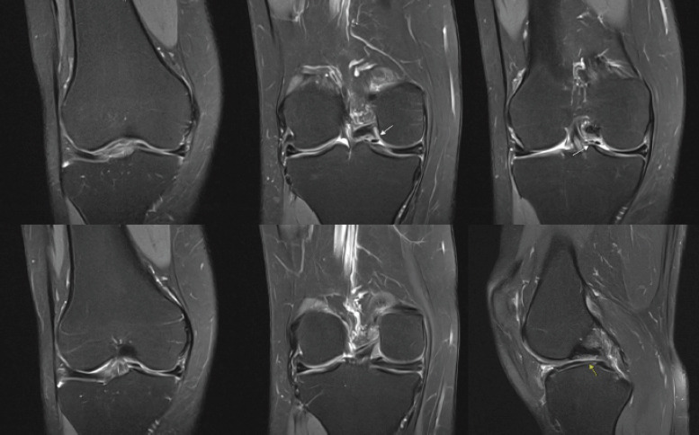
Introduction: Anomalous medial menisci are rare entities compared to their lateral counterparts. These anomalies include atypical insertions, most commonly into the anterior cruciate ligament, and discoid variants among others. This case adds to the literature on anomalous medial menisci with the presentation of a variant not described in the literature before.
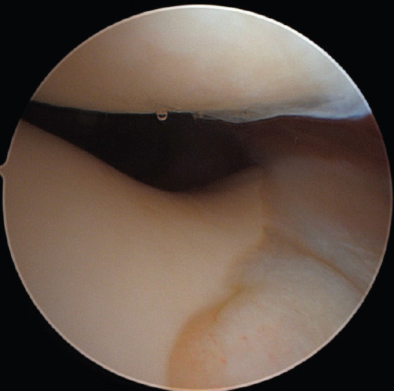
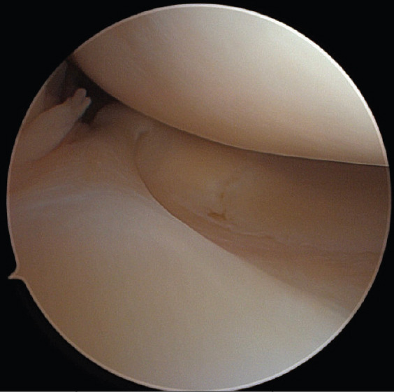
Case report: Our patient is a 38-year-old female who presented to the outpatient orthopedic clinic complaining of right knee pain in the setting of a traumatic kneeling event. She had a past medical history of asthma, polycystic ovary syndrome, and anxiety, but no surgical history related to the knee. She underwent magnetic resonance imaging identified what appeared to be a bucket-handle medial meniscus tear. The patient was brought to the operating room where diagnostic arthroscopy revealed a radial tear at the posterior horn of the medial meniscus as well as an anomalous connection between the anterior and posterior roots. The band specifically ran from the posterior horn of the medial meniscus, then superiorly and along the posterior cruciate ligament, and ultimately attached to the anterior horn of the medial meniscus in an "O" shape. In addition, a large patellofemoral plica was identified overlying the lateral femoral condyle. The tear, plica, and anomalous band were all debrided. The patient was progressing well on her most recent 7-month follow-up visit.
Conclusion: To our knowledge, this is the only reported case of such an anomaly. This case highlights the fact that there are likely other unidentified meniscal variants, and if they are not correctly identified on imaging, it could lead to patient mismanagement. Further research is needed into these variants.

 求助内容:
求助内容: 应助结果提醒方式:


