Sabare Naaharaaj, Mohammed Tavfiq, J K Giriraj Harshavardhan, Balaji Vijayan
{"title":"Chondroblastoma of the Right Distal Femur Medial Condyle: A Case Report.","authors":"Sabare Naaharaaj, Mohammed Tavfiq, J K Giriraj Harshavardhan, Balaji Vijayan","doi":"10.13107/jocr.2025.v15.i09.6074","DOIUrl":null,"url":null,"abstract":"<p><strong>Introduction: </strong>Chondroblastoma is a rare, benign but locally aggressive bone tumor typically affecting adolescents and young adults. It commonly arises in the epiphyseal region of long bones, particularly the distal femur. Despite its benign nature, it can cause significant joint dysfunction, pain, and disability. Early diagnosis is critical for preserving joint function and preventing recurrence.</p><p><strong>Case report: </strong>A 15-year-old female presented with progressive right knee pain of 6 months' duration, worsened by weight-bearing and minimally relieved by analgesics. Clinical examination revealed tenderness and immobile swelling in the region of the medial femoral condyle. Imaging showed characteristic \"chicken-wire\" calcification, and computed tomography-guided biopsy confirmed chondroblastoma. The patient underwent extended curettage, iliac crest bone grafting, and the use of synthetic bone substitutes. Post-operative rehabilitation showed a good recovery in range of motion and limb function, with no recurrence at follow-up.</p><p><strong>Conclusion: </strong>Early diagnosis and appropriate surgical management, including extended curettage and bone grafting, are essential to prevent recurrence and restore joint function in distal femur chondroblastoma. Functional outcomes are generally favorable with timely and targeted treatment.</p>","PeriodicalId":16647,"journal":{"name":"Journal of Orthopaedic Case Reports","volume":"15 9","pages":"213-218"},"PeriodicalIF":0.0000,"publicationDate":"2025-09-01","publicationTypes":"Journal Article","fieldsOfStudy":null,"isOpenAccess":false,"openAccessPdf":"https://www.ncbi.nlm.nih.gov/pmc/articles/PMC12422696/pdf/","citationCount":"0","resultStr":null,"platform":"Semanticscholar","paperid":null,"PeriodicalName":"Journal of Orthopaedic Case Reports","FirstCategoryId":"1085","ListUrlMain":"https://doi.org/10.13107/jocr.2025.v15.i09.6074","RegionNum":0,"RegionCategory":null,"ArticlePicture":[],"TitleCN":null,"AbstractTextCN":null,"PMCID":null,"EPubDate":"","PubModel":"","JCR":"","JCRName":"","Score":null,"Total":0}
引用次数: 0
Abstract
Introduction: Chondroblastoma is a rare, benign but locally aggressive bone tumor typically affecting adolescents and young adults. It commonly arises in the epiphyseal region of long bones, particularly the distal femur. Despite its benign nature, it can cause significant joint dysfunction, pain, and disability. Early diagnosis is critical for preserving joint function and preventing recurrence.
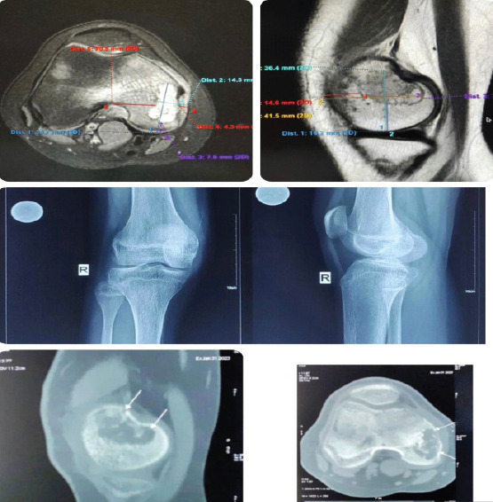
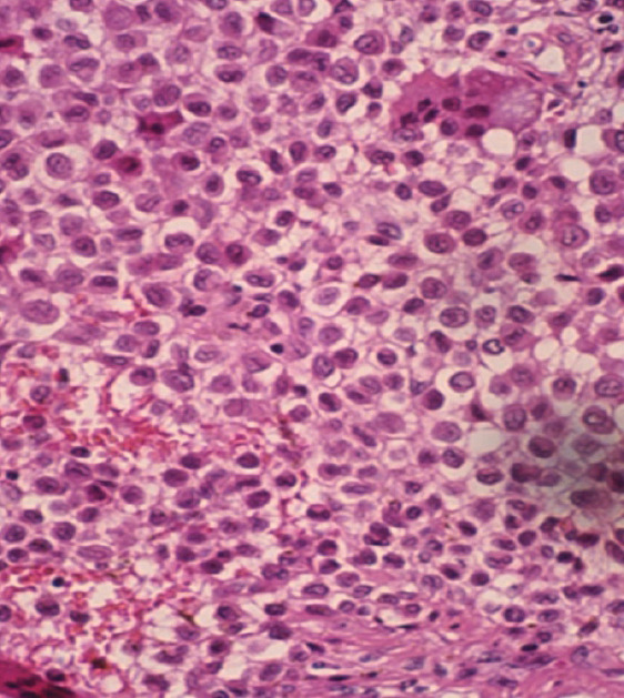
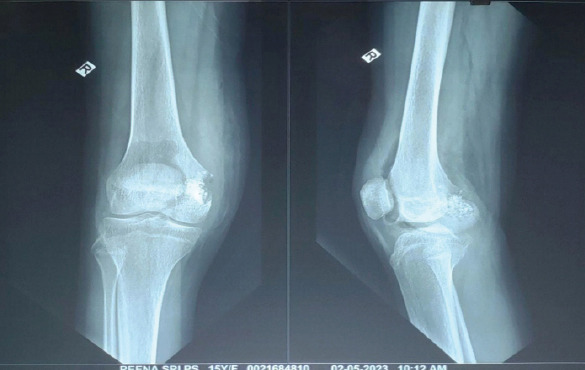
Case report: A 15-year-old female presented with progressive right knee pain of 6 months' duration, worsened by weight-bearing and minimally relieved by analgesics. Clinical examination revealed tenderness and immobile swelling in the region of the medial femoral condyle. Imaging showed characteristic "chicken-wire" calcification, and computed tomography-guided biopsy confirmed chondroblastoma. The patient underwent extended curettage, iliac crest bone grafting, and the use of synthetic bone substitutes. Post-operative rehabilitation showed a good recovery in range of motion and limb function, with no recurrence at follow-up.
Conclusion: Early diagnosis and appropriate surgical management, including extended curettage and bone grafting, are essential to prevent recurrence and restore joint function in distal femur chondroblastoma. Functional outcomes are generally favorable with timely and targeted treatment.

 求助内容:
求助内容: 应助结果提醒方式:


