Hong Yang, Fei Ma, Rui Li, Qiang Zhou, Fan Lin, Hesong Zeng, Dao Wen Wang, Jiangang Jiang, Xiang Luo, Hong Wang
{"title":"Subclinical Brain Lesions in Magnetic Resonance Imaging are a Potential Indicator of Patent Foramen Ovale Related Migraines in Younger Patients.","authors":"Hong Yang, Fei Ma, Rui Li, Qiang Zhou, Fan Lin, Hesong Zeng, Dao Wen Wang, Jiangang Jiang, Xiang Luo, Hong Wang","doi":"10.31083/RCM37480","DOIUrl":null,"url":null,"abstract":"<p><strong>Background: </strong>The causal relationship between migraines and patent foramen ovale (PFO) remains controversial, and a major unresolved question is how to define migraines attributable to PFO. Thus, this study aimed to determine if brain lesions could be a potential indicator of PFO-related migraines.</p><p><strong>Methods: </strong>Consecutive migraine patients from 2017 to 2019 who underwent transthoracic echocardiography or transcranial Doppler examination with an agitated saline contrast injection were assessed for right-to-left shunts. We then presented diffusion-weighted imaging (DWI) in brain magnetic resonance imaging and its association with PFO in the included patients.</p><p><strong>Results: </strong>A total of 424 patients with a mean age of 44.39 ± 12.06 years were included in this retrospective study. Among them, 244 patients (57.5%) had PFO, and 246 patients (58%) had subclinical brain lesions-the brain lesions presented as single or multiple scattered lesions. No association was observed between PFO prevalence and brain lesions in the total cohort (odds ratio (OR) 0.499); however, a significant association was observed in patients aged less than 46 years (OR, 3.614 in the group aged <34 years, 95% confidence interval (CI) 1.128-11.580, and 3.132 in the group of 34 years ≤ age < 46 years, 95% CI 1.334-7.350, respectively). Lesions in patients with PFO observed using DWI came more from the anterior or multiple than the posterior vascular territory (<i>p</i> = 0.033). DWI lesion numbers, location, and right-to-left shunt amounts did not affect the association between DWI-observed lesions and PFO.</p><p><strong>Conclusions: </strong>This study demonstrated that subclinical brain lesions are associated with PFO and may be used as a potential predictor of PFO-related migraines in patients aged less than 46 years. This may help identify candidate patients for PFO closure in future clinical decisions.</p>","PeriodicalId":20989,"journal":{"name":"Reviews in cardiovascular medicine","volume":"26 8","pages":"37480"},"PeriodicalIF":1.3000,"publicationDate":"2025-08-29","publicationTypes":"Journal Article","fieldsOfStudy":null,"isOpenAccess":false,"openAccessPdf":"https://www.ncbi.nlm.nih.gov/pmc/articles/PMC12415766/pdf/","citationCount":"0","resultStr":null,"platform":"Semanticscholar","paperid":null,"PeriodicalName":"Reviews in cardiovascular medicine","FirstCategoryId":"3","ListUrlMain":"https://doi.org/10.31083/RCM37480","RegionNum":4,"RegionCategory":"医学","ArticlePicture":[],"TitleCN":null,"AbstractTextCN":null,"PMCID":null,"EPubDate":"2025/8/1 0:00:00","PubModel":"eCollection","JCR":"Q3","JCRName":"CARDIAC & CARDIOVASCULAR SYSTEMS","Score":null,"Total":0}
引用次数: 0
Abstract
Background: The causal relationship between migraines and patent foramen ovale (PFO) remains controversial, and a major unresolved question is how to define migraines attributable to PFO. Thus, this study aimed to determine if brain lesions could be a potential indicator of PFO-related migraines.
Methods: Consecutive migraine patients from 2017 to 2019 who underwent transthoracic echocardiography or transcranial Doppler examination with an agitated saline contrast injection were assessed for right-to-left shunts. We then presented diffusion-weighted imaging (DWI) in brain magnetic resonance imaging and its association with PFO in the included patients.
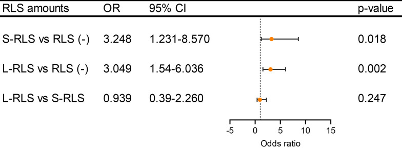
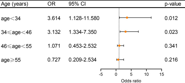
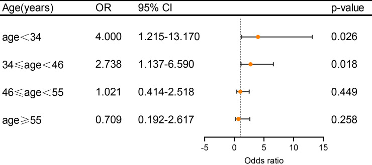
Results: A total of 424 patients with a mean age of 44.39 ± 12.06 years were included in this retrospective study. Among them, 244 patients (57.5%) had PFO, and 246 patients (58%) had subclinical brain lesions-the brain lesions presented as single or multiple scattered lesions. No association was observed between PFO prevalence and brain lesions in the total cohort (odds ratio (OR) 0.499); however, a significant association was observed in patients aged less than 46 years (OR, 3.614 in the group aged <34 years, 95% confidence interval (CI) 1.128-11.580, and 3.132 in the group of 34 years ≤ age < 46 years, 95% CI 1.334-7.350, respectively). Lesions in patients with PFO observed using DWI came more from the anterior or multiple than the posterior vascular territory (p = 0.033). DWI lesion numbers, location, and right-to-left shunt amounts did not affect the association between DWI-observed lesions and PFO.
Conclusions: This study demonstrated that subclinical brain lesions are associated with PFO and may be used as a potential predictor of PFO-related migraines in patients aged less than 46 years. This may help identify candidate patients for PFO closure in future clinical decisions.
期刊介绍:
RCM is an international, peer-reviewed, open access journal. RCM publishes research articles, review papers and short communications on cardiovascular medicine as well as research on cardiovascular disease. We aim to provide a forum for publishing papers which explore the pathogenesis and promote the progression of cardiac and vascular diseases. We also seek to establish an interdisciplinary platform, focusing on translational issues, to facilitate the advancement of research, clinical treatment and diagnostic procedures. Heart surgery, cardiovascular imaging, risk factors and various clinical cardiac & vascular research will be considered.
 求助内容:
求助内容: 应助结果提醒方式:


