Three-Dimensional Lower Extremity Alignment During the Stance Phase of Gait Using Anteroposterior Fluoroscopic Imaging and Image-Matching Technique: A Comparison with the Standing Position in Advanced Knee Osteoarthritis.
{"title":"Three-Dimensional Lower Extremity Alignment During the Stance Phase of Gait Using Anteroposterior Fluoroscopic Imaging and Image-Matching Technique: A Comparison with the Standing Position in Advanced Knee Osteoarthritis.","authors":"Tatsuya Soeno, Takashi Sato, Koichi Kobayashi, Ryota Katsumi, Kazutaka Otani, Hiroyuki Kawashima","doi":"10.2106/JBJS.OA.25.00168","DOIUrl":null,"url":null,"abstract":"<p><strong>Background: </strong>Lower extremity alignment in knee osteoarthritis (OA) is conventionally assessed using standing radiographs. However, symptoms often manifest during gait. Understanding dynamic alignment during gait may help characterize disease progression and inform treatment strategies.</p><p><strong>Methods: </strong>Twenty patients (40 knees) with advanced medial knee OA scheduled for arthroplasty were analyzed. Lower extremity alignment in standing (LEA-Standing) and during the midstance phase of gait (LEA-Gait) was evaluated using 3D-2D image matching technique with a ground-referenced and gravity-referenced coordinate system. Alignment parameters included femoral and tibial inclinations (coronal and sagittal), rotation angles, hip-knee-ankle angle (HKA), and tibial joint line angle (TJLA). Parameters were compared between gait and standing. ΔLEA (gait minus standing) was analyzed in relation with patient background and standing alignment. Medial joint space closure was evaluated on static radiographs and during gait.</p><p><strong>Results: </strong>LEA-Gait showed greater lateral inclination of the tibia and TJLA compared with standing, which resulted in increased varus HKA (all p < 0.01). No significant differences were observed in femoral or tibial rotation. Greater ΔTMA (tibial mechanical axis) and ΔTJLA were observed in knees with milder malalignment in standing. Medial joint space appeared open in 13 knees on standing and 5 on Rosenberg views but was closed in all 40 knees during gait.</p><p><strong>Conclusions: </strong>LEA-Gait differed significantly from LEA-Standing, revealing medial joint space closure and alignment abnormalities not captured by static evaluations including standing radiographs and Rosenberg views. These findings highlight the importance of considering the possibility of cartilage wear that may not be apparent on static radiographs.</p><p><strong>Level of evidence: </strong>Level II. See Instructions for Authors for a complete description of levels of evidence.</p>","PeriodicalId":36492,"journal":{"name":"JBJS Open Access","volume":"10 3","pages":""},"PeriodicalIF":3.8000,"publicationDate":"2025-09-08","publicationTypes":"Journal Article","fieldsOfStudy":null,"isOpenAccess":false,"openAccessPdf":"https://www.ncbi.nlm.nih.gov/pmc/articles/PMC12412737/pdf/","citationCount":"0","resultStr":null,"platform":"Semanticscholar","paperid":null,"PeriodicalName":"JBJS Open Access","FirstCategoryId":"1085","ListUrlMain":"https://doi.org/10.2106/JBJS.OA.25.00168","RegionNum":0,"RegionCategory":null,"ArticlePicture":[],"TitleCN":null,"AbstractTextCN":null,"PMCID":null,"EPubDate":"2025/7/1 0:00:00","PubModel":"eCollection","JCR":"Q2","JCRName":"ORTHOPEDICS","Score":null,"Total":0}
引用次数: 0
Abstract
Background: Lower extremity alignment in knee osteoarthritis (OA) is conventionally assessed using standing radiographs. However, symptoms often manifest during gait. Understanding dynamic alignment during gait may help characterize disease progression and inform treatment strategies.
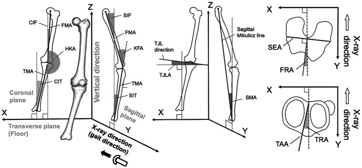
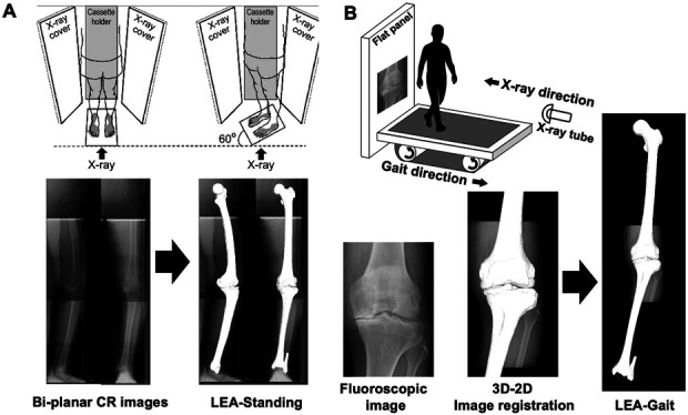
Methods: Twenty patients (40 knees) with advanced medial knee OA scheduled for arthroplasty were analyzed. Lower extremity alignment in standing (LEA-Standing) and during the midstance phase of gait (LEA-Gait) was evaluated using 3D-2D image matching technique with a ground-referenced and gravity-referenced coordinate system. Alignment parameters included femoral and tibial inclinations (coronal and sagittal), rotation angles, hip-knee-ankle angle (HKA), and tibial joint line angle (TJLA). Parameters were compared between gait and standing. ΔLEA (gait minus standing) was analyzed in relation with patient background and standing alignment. Medial joint space closure was evaluated on static radiographs and during gait.
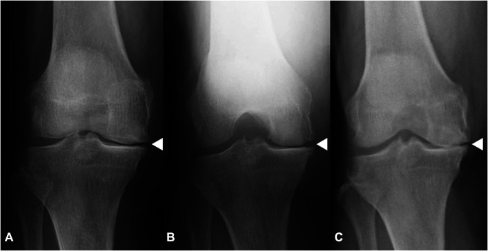
Results: LEA-Gait showed greater lateral inclination of the tibia and TJLA compared with standing, which resulted in increased varus HKA (all p < 0.01). No significant differences were observed in femoral or tibial rotation. Greater ΔTMA (tibial mechanical axis) and ΔTJLA were observed in knees with milder malalignment in standing. Medial joint space appeared open in 13 knees on standing and 5 on Rosenberg views but was closed in all 40 knees during gait.
Conclusions: LEA-Gait differed significantly from LEA-Standing, revealing medial joint space closure and alignment abnormalities not captured by static evaluations including standing radiographs and Rosenberg views. These findings highlight the importance of considering the possibility of cartilage wear that may not be apparent on static radiographs.
Level of evidence: Level II. See Instructions for Authors for a complete description of levels of evidence.

 求助内容:
求助内容: 应助结果提醒方式:


