Maxillofacial growth retarded by microsurgical injuries in young rats
引用次数: 0
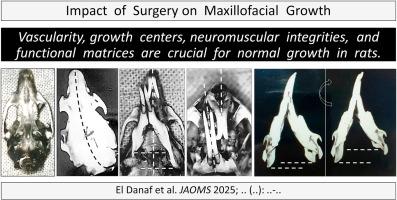
Abstract
This study aimed to experimentally evaluate the impact of maxillofacial surgically induced trauma on the growth of the skull and facial bones. The materials were previously divided into two separate papers and published in French; however, this research article has assembled the methods and results, added measurable traces to figures, and updated the discussion, references, and layout.
Methods
Twelve maxillofacial injuries were tempted in 100 rats at the age of 8 ± 2 days. Surgeries were performed under surgical microscope to precisely hit a specific target. Sixty-nine rats survived to the age of 36 ± 2 days and were sacrificed. The morphology and bilateral dimensions of dry skull and facial skeleton in each group were compared with those of ten rats that escaped surgery and used as controls.
Results
Seven procedures including coagulation of palatine blood supply, excision of the mid-palate sagittal suture, removal of a mandibular condyle, disinsertion, or excision of a masseter muscle, cutting of a mandibular nerve, and enucleation of an eye globe, led to a deterioration of facial skeleton growth. However, five procedures including palatal subperiosteal dissection, mucoperiosteal excision, unilateral nasal obliteration, external carotid artery ligation, and facial nerve interruption did not hinder growth.
Conclusion
Preserving the integrity of vascularity, periosteal osteogenic power, sutures, condyle growth centers, masticatory neuromuscular function, and orbit/globe capsular matrix, are crucial for normal craniofacial growth and development.
显微外科损伤对幼鼠颌面部发育的影响
本研究旨在通过实验评估颌面外科创伤对颅骨和面骨生长的影响。这些材料以前被分成两篇单独的论文,用法语出版;然而,本文整合了方法和结果,在图中增加了可测量的痕迹,并更新了讨论、参考文献和布局。方法采用8±2日龄大鼠100只,引诱12只颌面部损伤。手术在手术显微镜下进行,以精确地击中特定的目标。69只大鼠存活至36±2天,处死。各组干颅骨和面部骨骼的形态学和双侧尺寸与10只手术逃脱的大鼠作为对照进行比较。结果包括腭血供凝固、切除中腭矢状缝合线、移除下颌髁突、拔出或切除咬肌、切断下颌神经、摘除眼球等7项手术导致面部骨骼生长恶化。然而,包括腭骨膜下剥离、粘骨膜切除、单侧鼻闭塞、颈外动脉结扎和面神经中断在内的五种手术并未阻碍生长。结论保留血管、骨膜成骨力、缝合线、髁突生长中心、咀嚼神经肌肉功能和眶/球囊基质的完整性是颅面正常生长发育的关键。
本文章由计算机程序翻译,如有差异,请以英文原文为准。
求助全文
约1分钟内获得全文
求助全文

 求助内容:
求助内容: 应助结果提醒方式:


