Evaluating endometrial response to human chorionic gonadotropin: alterations in epigenetic regulation and extracellular vesicle cargo of endometrial stromal cells.
{"title":"Evaluating endometrial response to human chorionic gonadotropin: alterations in epigenetic regulation and extracellular vesicle cargo of endometrial stromal cells.","authors":"Deimantė Žukauskaitė, Erika Girniūtė, Rūta Navakauskienė","doi":"10.1093/hropen/hoaf051","DOIUrl":null,"url":null,"abstract":"<p><strong>Study question: </strong>What is the effect of hCG on the epigenetic profile and the expression of other molecular factors in endometrial stromal cells (ESCs)?</p><p><strong>Summary answer: </strong>Our findings suggest that hCG treatment alters the molecular environment of decidualized ESCs, potentially influencing implantation and immune regulation through epigenetic modifications and changes in the levels of secreted proteins and micro-ribonucleic acids (miRNAs).</p><p><strong>What is known already: </strong>Embryo implantation depends not only on the quality of the embryo but also on the receptivity of the endometrium, the specialized lining of the uterus that undergoes dynamic changes to support pregnancy. Effective communication between the maternal and fetal compartments, facilitated by molecular signals and cellular interactions, is essential for successful implantation.</p><p><strong>Study design size duration: </strong>Cross-sectional study of patient-derived ESCs comparing untreated cells with cells treated with hCG and/or decidualization induction. The number of samples depends on the method and varies from 2 to 8. Results were analyzed after 6-, 24-, 48-, and 72-h time-points.</p><p><strong>Participants/materials setting methods: </strong>ESCs were isolated from patients undergoing assisted reproductive technologies. In the study, we analyzed changes in the epigenetic profile and other molecular factors of ESCs during decidualization and in <i>in vitro</i> response to the embryo-secreted factor, hCG. ESCs were induced for decidualization for 3 days (medroxyprogesterone acetate+cAMP), or treated with hCG for 24 h, or given combined treatment: 2 days of decidualization followed by 24 h of hCG. Furthermore, we compared decidualized ESCs with decidualized ESCs that were also treated with hCG. We examined various cellular properties, including morphology, metabolic activity, and cell viability of ESCs after induction of decidualization and hCG treatment. Additionally, we assessed changes in the expression of genes associated with decidualization, inflammatory response, apoptosis regulation, and epigenetic factors using RT-qPCR. The levels of histone modifications and the factors regulating these modifications were explored by performing western blot assays. Additionally, we performed a chromatin immunoprecipitation assay to extract gene regions enriched with the epigenetic modification H3K27Ac. Finally, we analyzed the protein and miRNA level changes in ESC extracellular vesicles (ESC-EVs) after the indicated treatments, using mass spectrometry and small RNA sequencing.</p><p><strong>Main results and the role of chance: </strong>Our study found that hCG treatment increased prolactin gene (<i>PRL)</i> expression (<i>P</i> < 0.05), while the expression of <i>IL6</i> and <i>BAK1</i> was inhibited in ESCs (<i>P</i> < 0.05). We also revealed that hCG affects epigenetic regulation, leading to changes in the expression of <i>EED</i>, <i>HDAC1</i>, and <i>TET1/2/3</i> (<i>P</i> < 0.05). Specifically, hCG treatment resulted in increased levels of H3K27Ac in gene regions associated with decidualization, such as <i>FOXO1</i> (<i>P</i> < 0.05), and implantation genes like <i>HOXA10</i> and <i>HAND2</i> (<i>P</i> = 0.06). After decidualization, we observed increased protein levels in ESC-EVs that are associated with embryo implantation (<i>P</i> = 0.0038) and pregnancy (<i>P</i> = 0.0012). These included proteins such as FIBL-1, IGFBP-1/7, MMP-2, STC-1/2, and PAPP-A (<i>P</i> < 0.05). Additionally, hCG treatment in decidualized ESCs elevated the levels of proteins involved in immune system regulation, including PR-3 (<i>P</i> = 0.0131). Moreover, we revealed changes in miRNA levels within EVs secreted by ESCs following hCG treatment and decidualization, which are associated with embryo development in the uterus (<i>P</i> = 0.03). Notable miRNAs include hsa-miR-340-3p, hsa-miR-663a, hsa-miR-766-5p, hsa-miR-3138, and hsa-miR-3180-5p (<i>P</i> < 0.05).</p><p><strong>Large scale data: </strong>N/A.</p><p><strong>Limitations reasons for caution: </strong>This study utilizes a comprehensive analysis to explore potential epigenetic and molecular targets essential for endometrial function. The research is based on <i>in vitro</i> experiments using ESCs derived from <i>ex vivo</i> samples. Future studies involving a broader range of cell types and larger sample sizes could help to further validate and expand upon these results.</p><p><strong>Wider implications of the findings: </strong>We found that hCG enhances the decidualization process in a dose-dependent manner and affects implantation and immune regulation through epigenetic changes, as well as variations in the levels of secreted proteins and miRNAs. Our study suggests that the application of hCG in assisted reproduction technologies may offer potential benefits for patients. However, carefully considering the appropriate dosage is important to ensure optimal outcomes.</p><p><strong>Study funding/competing interests: </strong>This research did not receive any specific grant from funding agencies in the public, commercial, or not-for-profit sectors. The authors have no conflicts of interest to declare.</p>","PeriodicalId":73264,"journal":{"name":"Human reproduction open","volume":"2025 3","pages":"hoaf051"},"PeriodicalIF":11.1000,"publicationDate":"2025-08-14","publicationTypes":"Journal Article","fieldsOfStudy":null,"isOpenAccess":false,"openAccessPdf":"https://www.ncbi.nlm.nih.gov/pmc/articles/PMC12408481/pdf/","citationCount":"0","resultStr":null,"platform":"Semanticscholar","paperid":null,"PeriodicalName":"Human reproduction open","FirstCategoryId":"1085","ListUrlMain":"https://doi.org/10.1093/hropen/hoaf051","RegionNum":0,"RegionCategory":null,"ArticlePicture":[],"TitleCN":null,"AbstractTextCN":null,"PMCID":null,"EPubDate":"2025/1/1 0:00:00","PubModel":"eCollection","JCR":"Q1","JCRName":"OBSTETRICS & GYNECOLOGY","Score":null,"Total":0}
引用次数: 0
Abstract
Study question: What is the effect of hCG on the epigenetic profile and the expression of other molecular factors in endometrial stromal cells (ESCs)?
Summary answer: Our findings suggest that hCG treatment alters the molecular environment of decidualized ESCs, potentially influencing implantation and immune regulation through epigenetic modifications and changes in the levels of secreted proteins and micro-ribonucleic acids (miRNAs).
What is known already: Embryo implantation depends not only on the quality of the embryo but also on the receptivity of the endometrium, the specialized lining of the uterus that undergoes dynamic changes to support pregnancy. Effective communication between the maternal and fetal compartments, facilitated by molecular signals and cellular interactions, is essential for successful implantation.
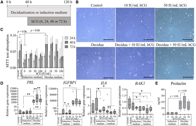
Study design size duration: Cross-sectional study of patient-derived ESCs comparing untreated cells with cells treated with hCG and/or decidualization induction. The number of samples depends on the method and varies from 2 to 8. Results were analyzed after 6-, 24-, 48-, and 72-h time-points.
Participants/materials setting methods: ESCs were isolated from patients undergoing assisted reproductive technologies. In the study, we analyzed changes in the epigenetic profile and other molecular factors of ESCs during decidualization and in in vitro response to the embryo-secreted factor, hCG. ESCs were induced for decidualization for 3 days (medroxyprogesterone acetate+cAMP), or treated with hCG for 24 h, or given combined treatment: 2 days of decidualization followed by 24 h of hCG. Furthermore, we compared decidualized ESCs with decidualized ESCs that were also treated with hCG. We examined various cellular properties, including morphology, metabolic activity, and cell viability of ESCs after induction of decidualization and hCG treatment. Additionally, we assessed changes in the expression of genes associated with decidualization, inflammatory response, apoptosis regulation, and epigenetic factors using RT-qPCR. The levels of histone modifications and the factors regulating these modifications were explored by performing western blot assays. Additionally, we performed a chromatin immunoprecipitation assay to extract gene regions enriched with the epigenetic modification H3K27Ac. Finally, we analyzed the protein and miRNA level changes in ESC extracellular vesicles (ESC-EVs) after the indicated treatments, using mass spectrometry and small RNA sequencing.
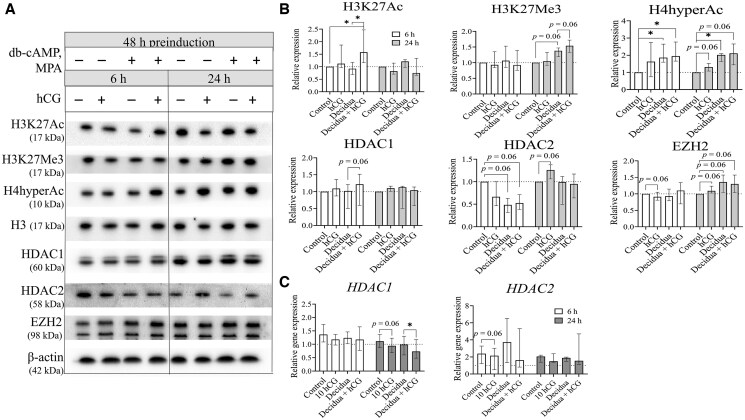
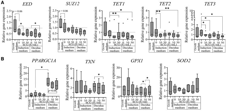
Main results and the role of chance: Our study found that hCG treatment increased prolactin gene (PRL) expression (P < 0.05), while the expression of IL6 and BAK1 was inhibited in ESCs (P < 0.05). We also revealed that hCG affects epigenetic regulation, leading to changes in the expression of EED, HDAC1, and TET1/2/3 (P < 0.05). Specifically, hCG treatment resulted in increased levels of H3K27Ac in gene regions associated with decidualization, such as FOXO1 (P < 0.05), and implantation genes like HOXA10 and HAND2 (P = 0.06). After decidualization, we observed increased protein levels in ESC-EVs that are associated with embryo implantation (P = 0.0038) and pregnancy (P = 0.0012). These included proteins such as FIBL-1, IGFBP-1/7, MMP-2, STC-1/2, and PAPP-A (P < 0.05). Additionally, hCG treatment in decidualized ESCs elevated the levels of proteins involved in immune system regulation, including PR-3 (P = 0.0131). Moreover, we revealed changes in miRNA levels within EVs secreted by ESCs following hCG treatment and decidualization, which are associated with embryo development in the uterus (P = 0.03). Notable miRNAs include hsa-miR-340-3p, hsa-miR-663a, hsa-miR-766-5p, hsa-miR-3138, and hsa-miR-3180-5p (P < 0.05).
Large scale data: N/A.
Limitations reasons for caution: This study utilizes a comprehensive analysis to explore potential epigenetic and molecular targets essential for endometrial function. The research is based on in vitro experiments using ESCs derived from ex vivo samples. Future studies involving a broader range of cell types and larger sample sizes could help to further validate and expand upon these results.
Wider implications of the findings: We found that hCG enhances the decidualization process in a dose-dependent manner and affects implantation and immune regulation through epigenetic changes, as well as variations in the levels of secreted proteins and miRNAs. Our study suggests that the application of hCG in assisted reproduction technologies may offer potential benefits for patients. However, carefully considering the appropriate dosage is important to ensure optimal outcomes.
Study funding/competing interests: This research did not receive any specific grant from funding agencies in the public, commercial, or not-for-profit sectors. The authors have no conflicts of interest to declare.

 求助内容:
求助内容: 应助结果提醒方式:


