The role of low subcortical iron, white matter myelin, and oligodendrocytes in schizophrenia: a quantitative susceptibility mapping and diffusion tensor imaging study
Luke J. Vano, Robert A. McCutcheon, Jan Sedlacik, Grazia Rutigliano, Stephen J. Kaar, Valeria Finelli, Maria C. Lobo, Alaine Berry, Ben Statton, Amir Fazlollahi, Ian P. Everall, Oliver D. Howes
{"title":"The role of low subcortical iron, white matter myelin, and oligodendrocytes in schizophrenia: a quantitative susceptibility mapping and diffusion tensor imaging study","authors":"Luke J. Vano, Robert A. McCutcheon, Jan Sedlacik, Grazia Rutigliano, Stephen J. Kaar, Valeria Finelli, Maria C. Lobo, Alaine Berry, Ben Statton, Amir Fazlollahi, Ian P. Everall, Oliver D. Howes","doi":"10.1038/s41380-025-03195-7","DOIUrl":null,"url":null,"abstract":"<p>Iron—the most abundant magnetic brain substance—is essential for many biological processes, including dopamine and myelin synthesis. Quantitative susceptibility mapping (QSM) MRI has recently linked altered subcortical magnetic susceptibility (χ) to schizophrenia. Since χ is increased by iron and decreased by myelin, abnormal levels of either could underlie these QSM differences. In white matter tracts, magnetic susceptibility anisotropy (δχ) serves as a myelin-specific marker that is insensitive to iron content. To clarify the origin of case-control χ differences, we employed QSM in 85 individuals with schizophrenia, from first-episode mental health teams, and 86 healthy controls. A subset also underwent diffusion tensor imaging (DTI) to calculate subcortical tissue mean diffusivity, which inversely correlates with myelin concentration and fractional anisotropy. White matter δχ was calculated by combining QSM and DTI. Schizophrenia was associated with lower subcortical χ (d = −0.36, p = 0.023). This was significant in the caudate nucleus (d = −0.37, p = 0.037), putamen (d = −0.36, p = 0.037), globus pallidus (d = −0.57, p = 0.001), and SN-VTA (as previously reported). Additionally, schizophrenia was linked to higher subcortical mean diffusivity (d = 0.44, p = 0.018), and lower white matter δχ (d = −0.37, p = 0.047). These findings suggest that both subcortical iron and brain myelin levels are lower in schizophrenia. By comparing our voxelwise χ maps with postmortem gene expression data, we reveal that regions with lower subcortical χ in schizophrenia are enriched for oligodendrocyte-related genes (p < 0.001). As oligodendrocytes are both the most iron-rich brain cells and essential for myelin synthesis, our results implicate oligodendrocyte dysfunction in schizophrenia pathophysiology.</p>","PeriodicalId":19008,"journal":{"name":"Molecular Psychiatry","volume":"48 1","pages":""},"PeriodicalIF":10.1000,"publicationDate":"2025-09-05","publicationTypes":"Journal Article","fieldsOfStudy":null,"isOpenAccess":false,"openAccessPdf":"","citationCount":"0","resultStr":null,"platform":"Semanticscholar","paperid":null,"PeriodicalName":"Molecular Psychiatry","FirstCategoryId":"3","ListUrlMain":"https://doi.org/10.1038/s41380-025-03195-7","RegionNum":1,"RegionCategory":"医学","ArticlePicture":[],"TitleCN":null,"AbstractTextCN":null,"PMCID":null,"EPubDate":"","PubModel":"","JCR":"Q1","JCRName":"BIOCHEMISTRY & MOLECULAR BIOLOGY","Score":null,"Total":0}
引用次数: 0
Abstract
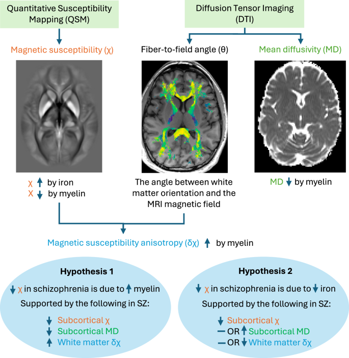
Iron—the most abundant magnetic brain substance—is essential for many biological processes, including dopamine and myelin synthesis. Quantitative susceptibility mapping (QSM) MRI has recently linked altered subcortical magnetic susceptibility (χ) to schizophrenia. Since χ is increased by iron and decreased by myelin, abnormal levels of either could underlie these QSM differences. In white matter tracts, magnetic susceptibility anisotropy (δχ) serves as a myelin-specific marker that is insensitive to iron content. To clarify the origin of case-control χ differences, we employed QSM in 85 individuals with schizophrenia, from first-episode mental health teams, and 86 healthy controls. A subset also underwent diffusion tensor imaging (DTI) to calculate subcortical tissue mean diffusivity, which inversely correlates with myelin concentration and fractional anisotropy. White matter δχ was calculated by combining QSM and DTI. Schizophrenia was associated with lower subcortical χ (d = −0.36, p = 0.023). This was significant in the caudate nucleus (d = −0.37, p = 0.037), putamen (d = −0.36, p = 0.037), globus pallidus (d = −0.57, p = 0.001), and SN-VTA (as previously reported). Additionally, schizophrenia was linked to higher subcortical mean diffusivity (d = 0.44, p = 0.018), and lower white matter δχ (d = −0.37, p = 0.047). These findings suggest that both subcortical iron and brain myelin levels are lower in schizophrenia. By comparing our voxelwise χ maps with postmortem gene expression data, we reveal that regions with lower subcortical χ in schizophrenia are enriched for oligodendrocyte-related genes (p < 0.001). As oligodendrocytes are both the most iron-rich brain cells and essential for myelin synthesis, our results implicate oligodendrocyte dysfunction in schizophrenia pathophysiology.
期刊介绍:
Molecular Psychiatry focuses on publishing research that aims to uncover the biological mechanisms behind psychiatric disorders and their treatment. The journal emphasizes studies that bridge pre-clinical and clinical research, covering cellular, molecular, integrative, clinical, imaging, and psychopharmacology levels.
 求助内容:
求助内容: 应助结果提醒方式:


