{"title":"[A protein-specific quantitative detection method based on polyacrylamide gel electrophoresis and online fluorescence imaging].","authors":"Rui Zou, Zi-Xian Yu, Ze-Hua Guo, Hao-Zheng Dai, Qiang Zhang, Wei-Wen Liu, Cheng-Xi Cao","doi":"10.3724/SP.J.1123.2024.12017","DOIUrl":null,"url":null,"abstract":"<p><p>Specific protein detection plays a crucial role in biological analysis and clinical diagnostics, serving as an essential tool for disease diagnosis, therapeutic monitoring, and biological research. However, conventional methods such as immunofixation electrophoresis (IFE) and western blotting (WB) suffer from complex workflows, time-consuming operations, and limited quantification capabilities owing to intricate staining and de-staining procedures. In addition, these traditional immunological detection methods require extensive manual handling and specialized expertise, while low levels of automation restrict their applicability to high-throughput or large-scale analysis scenarios. Moreover, the multistep nature of these methods increases the risk of experimental errors and compromises quantification accuracy.Herein, we present a quantitative protein immune polyacrylamide gel electrophoresis (PAGE) detection method that combines immune-recognition principles with online fluorescence imaging technology, thereby offering a rapid and specific approach for quantifying target proteins. The developed method exploits the specificity of fluorescently labeled antibodies and the separation capability of PAGE, with formaldehyde crosslinking used to stabilize antigen-antibody complexes under the denaturing conditions of electrophoresis, thereby ensuring reliable quantification. The entire experimental workflow can be completed within 1.5 h and consists of three main steps. Firstly, the target protein is incubated with fluorescently labeled antibodies at room temperature for 0.5 h to form immune complexes, after which they are crosslinked using formaldehyde. The cross-linked samples are then loaded onto polyacrylamide gels and separated under optimized electrophoresis conditions (120 V, 15 min; 150 V, 15 min; 200 V, 15 min). Electrophoretic separation is finally monitored in real-time using an online fluorescence imaging system, which enables direct visualization of protein migration and eliminates the need for post-separation processing. Three distinct bands are observed on the precast gel following immune PAGE separation: the immune complexes at the uppermost position, the free fluorescent antibodies in the middle, and other proteins at the bottom. ImageJ software is used to analyze the electrophoresis pattern, and quantification is achieved based on the linear relationship between the fluorescence intensity of the free antibody and the mass concentration of the target protein. We systematically validated the performance of the method using human transferrin (TRF) as the model antigen protein and fluorescein-isothiocyanate-labeled (FITC-labeled) anti-TRF IgG antibody (anti-TRF IgG-FITC) as the detection probe, which involved analyzing three key aspects: the necessity of the formaldehyde-crosslinking step for maintaining immune complex stability, antibody recognition specificity in complex samples, and the linear correlation between the fluorescence signal and the mass concentration of the target protein. The method exhibited excellent analytical performance, with a linear range that extended between 5.0 and 200.0 mg/L and a correlation coefficient (<i>R</i><sup>2</sup>) of 0.993 0. Triplicate measurements of fluorescence intensity at all mass concentration points revealed a maximum relative standard deviation (RSD) of 1.65%. The limit of detection (LOD) reaches 0.5 mg/L, with recoveries between 98.2% and 105.0%. Repeatability experiments revealed maximum intra- and inter-day RSDs of 1.21% and 1.58%, respectively. Specificity testing confirmed that the developed method accurately quantified TRF without interference from other proteins in complex samples. These results highlight the good accuracy, excellent consistency, high sensitivity, and robust specificity of the developed method, thereby confirming its reliability for use in precise protein-quantification applications. Compared to traditional PAGE methods, the immune PAGE method introduced herein provides the ability to selectively quantify specific target proteins online using real-time fluorescence imaging technology. The method exhibits several notable advantages, including high resolution, a simple workflow, strong specificity, rapid analysis, and good reproducibility. The online fluorescence imaging system eliminates the need for complex gel-dismantling, membrane-transfer, fixation, staining, and de-staining steps while effectively preventing protein band broadening, thereby enabling highly sensitive quantitative analyses with superior resolution. The cost-effectiveness of the developed method is achieved through economical fluorescently labeled antibodies and low sample consumption. Moreover, the fundamental principle of the immune PAGE method suggests that this approach is readily adaptable to the quantification of other proteins through the judicious selection of specific fluorescently labeled antibodies. Consequently, the versatility of the developed method makes it a comprehensive analytical platform suitable for pharmaceutical preparations and clinical diagnostics, in which rapid and accurate protein quantification is essential for decision-making processes. Additionally, this method is an ideal choice for high-throughput applications in both academic research and industrial settings owing to the integration of automated analysis and short operation times. The protein immune PAGE method represents a significant advancement in specific protein quantification methodology with great potential as a promising tool that is expected to be widely adopted in various biological analysis scenarios.</p>","PeriodicalId":101336,"journal":{"name":"Se pu = Chinese journal of chromatography","volume":"43 9","pages":"1070-1077"},"PeriodicalIF":0.0000,"publicationDate":"2025-09-01","publicationTypes":"Journal Article","fieldsOfStudy":null,"isOpenAccess":false,"openAccessPdf":"https://www.ncbi.nlm.nih.gov/pmc/articles/PMC12412021/pdf/","citationCount":"0","resultStr":null,"platform":"Semanticscholar","paperid":null,"PeriodicalName":"Se pu = Chinese journal of chromatography","FirstCategoryId":"1085","ListUrlMain":"https://doi.org/10.3724/SP.J.1123.2024.12017","RegionNum":0,"RegionCategory":null,"ArticlePicture":[],"TitleCN":null,"AbstractTextCN":null,"PMCID":null,"EPubDate":"","PubModel":"","JCR":"","JCRName":"","Score":null,"Total":0}
引用次数: 0
Abstract
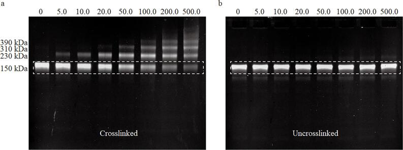
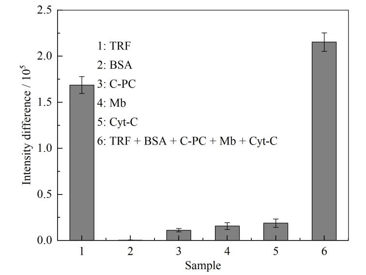
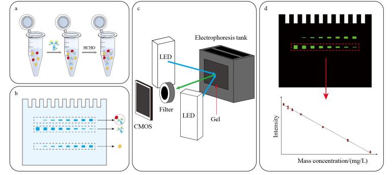
Specific protein detection plays a crucial role in biological analysis and clinical diagnostics, serving as an essential tool for disease diagnosis, therapeutic monitoring, and biological research. However, conventional methods such as immunofixation electrophoresis (IFE) and western blotting (WB) suffer from complex workflows, time-consuming operations, and limited quantification capabilities owing to intricate staining and de-staining procedures. In addition, these traditional immunological detection methods require extensive manual handling and specialized expertise, while low levels of automation restrict their applicability to high-throughput or large-scale analysis scenarios. Moreover, the multistep nature of these methods increases the risk of experimental errors and compromises quantification accuracy.Herein, we present a quantitative protein immune polyacrylamide gel electrophoresis (PAGE) detection method that combines immune-recognition principles with online fluorescence imaging technology, thereby offering a rapid and specific approach for quantifying target proteins. The developed method exploits the specificity of fluorescently labeled antibodies and the separation capability of PAGE, with formaldehyde crosslinking used to stabilize antigen-antibody complexes under the denaturing conditions of electrophoresis, thereby ensuring reliable quantification. The entire experimental workflow can be completed within 1.5 h and consists of three main steps. Firstly, the target protein is incubated with fluorescently labeled antibodies at room temperature for 0.5 h to form immune complexes, after which they are crosslinked using formaldehyde. The cross-linked samples are then loaded onto polyacrylamide gels and separated under optimized electrophoresis conditions (120 V, 15 min; 150 V, 15 min; 200 V, 15 min). Electrophoretic separation is finally monitored in real-time using an online fluorescence imaging system, which enables direct visualization of protein migration and eliminates the need for post-separation processing. Three distinct bands are observed on the precast gel following immune PAGE separation: the immune complexes at the uppermost position, the free fluorescent antibodies in the middle, and other proteins at the bottom. ImageJ software is used to analyze the electrophoresis pattern, and quantification is achieved based on the linear relationship between the fluorescence intensity of the free antibody and the mass concentration of the target protein. We systematically validated the performance of the method using human transferrin (TRF) as the model antigen protein and fluorescein-isothiocyanate-labeled (FITC-labeled) anti-TRF IgG antibody (anti-TRF IgG-FITC) as the detection probe, which involved analyzing three key aspects: the necessity of the formaldehyde-crosslinking step for maintaining immune complex stability, antibody recognition specificity in complex samples, and the linear correlation between the fluorescence signal and the mass concentration of the target protein. The method exhibited excellent analytical performance, with a linear range that extended between 5.0 and 200.0 mg/L and a correlation coefficient (R2) of 0.993 0. Triplicate measurements of fluorescence intensity at all mass concentration points revealed a maximum relative standard deviation (RSD) of 1.65%. The limit of detection (LOD) reaches 0.5 mg/L, with recoveries between 98.2% and 105.0%. Repeatability experiments revealed maximum intra- and inter-day RSDs of 1.21% and 1.58%, respectively. Specificity testing confirmed that the developed method accurately quantified TRF without interference from other proteins in complex samples. These results highlight the good accuracy, excellent consistency, high sensitivity, and robust specificity of the developed method, thereby confirming its reliability for use in precise protein-quantification applications. Compared to traditional PAGE methods, the immune PAGE method introduced herein provides the ability to selectively quantify specific target proteins online using real-time fluorescence imaging technology. The method exhibits several notable advantages, including high resolution, a simple workflow, strong specificity, rapid analysis, and good reproducibility. The online fluorescence imaging system eliminates the need for complex gel-dismantling, membrane-transfer, fixation, staining, and de-staining steps while effectively preventing protein band broadening, thereby enabling highly sensitive quantitative analyses with superior resolution. The cost-effectiveness of the developed method is achieved through economical fluorescently labeled antibodies and low sample consumption. Moreover, the fundamental principle of the immune PAGE method suggests that this approach is readily adaptable to the quantification of other proteins through the judicious selection of specific fluorescently labeled antibodies. Consequently, the versatility of the developed method makes it a comprehensive analytical platform suitable for pharmaceutical preparations and clinical diagnostics, in which rapid and accurate protein quantification is essential for decision-making processes. Additionally, this method is an ideal choice for high-throughput applications in both academic research and industrial settings owing to the integration of automated analysis and short operation times. The protein immune PAGE method represents a significant advancement in specific protein quantification methodology with great potential as a promising tool that is expected to be widely adopted in various biological analysis scenarios.

 求助内容:
求助内容: 应助结果提醒方式:


