Gerard R Hall, Sarah J Gascoigne, Jonathan J Horsley, Yujiang Wang, Csaba Kozma, Jane de Tisi, Sjoerd B Vos, Gavin P Winston, John S Duncan, Peter N Taylor
{"title":"Superficial and deep white matter abnormalities in temporal lobe epilepsy.","authors":"Gerard R Hall, Sarah J Gascoigne, Jonathan J Horsley, Yujiang Wang, Csaba Kozma, Jane de Tisi, Sjoerd B Vos, Gavin P Winston, John S Duncan, Peter N Taylor","doi":"10.1093/braincomms/fcaf305","DOIUrl":null,"url":null,"abstract":"<p><p>Non-invasive neuroimaging is important in epilepsy to help identify cerebral abnormalities. Abnormally reduced fractional anisotropy (FA) in deep white matter (WM) from diffusion-weighted imaging (DWI) is widely reported in large multi-cohort studies across all types of epilepsies. However, abnormalities in FA for superficial WM are rarely investigated in epilepsy. To gain a greater understanding of the nature of WM abnormality at different WM depths, we investigated DWI abnormalities at a range of superficial and deep WM in two separate temporal lobe epilepsy (TLE) cohorts. The first cohort (TLE = 81, Healthy Control; HC = 67) underwent a high angular resolution multi-shell DWI, whilst the second cohort (TLE = 70, HC = 29) had a single-shell acquisition. We registered FA maps to a standard template, and analysed temporal WM within 8 mm of the temporal lobe grey matter, amygdala and hippocampus. We standardised FA measures at different depths, and compared ipsi-versus contralateral temporal WM, and MRI-positive versus MRI-negative groups. We report three major findings: First, superficial WM had greater FA reductions than deep WM in TLE (<i>P</i> < 0.001). Second, this effect was more prominent in the ipsilateral than contralateral temporal lobe WM (<i>P</i> < 0.001). Third, these effects were present to a similar degree in patients who reported an MRI negative. All results are held in both TLE cohorts. These findings suggest that, in the temporal lobe, superficial WM is more abnormal than deep WM in TLE, with potential clinical use for lateralisation even in MRI-negative patients. These findings motivate further investigation of the importance of superficial WM in epilepsy.</p>","PeriodicalId":93915,"journal":{"name":"Brain communications","volume":"7 5","pages":"fcaf305"},"PeriodicalIF":4.5000,"publicationDate":"2025-08-19","publicationTypes":"Journal Article","fieldsOfStudy":null,"isOpenAccess":false,"openAccessPdf":"https://www.ncbi.nlm.nih.gov/pmc/articles/PMC12402771/pdf/","citationCount":"0","resultStr":null,"platform":"Semanticscholar","paperid":null,"PeriodicalName":"Brain communications","FirstCategoryId":"1085","ListUrlMain":"https://doi.org/10.1093/braincomms/fcaf305","RegionNum":0,"RegionCategory":null,"ArticlePicture":[],"TitleCN":null,"AbstractTextCN":null,"PMCID":null,"EPubDate":"2025/1/1 0:00:00","PubModel":"eCollection","JCR":"Q1","JCRName":"CLINICAL NEUROLOGY","Score":null,"Total":0}
引用次数: 0
Abstract
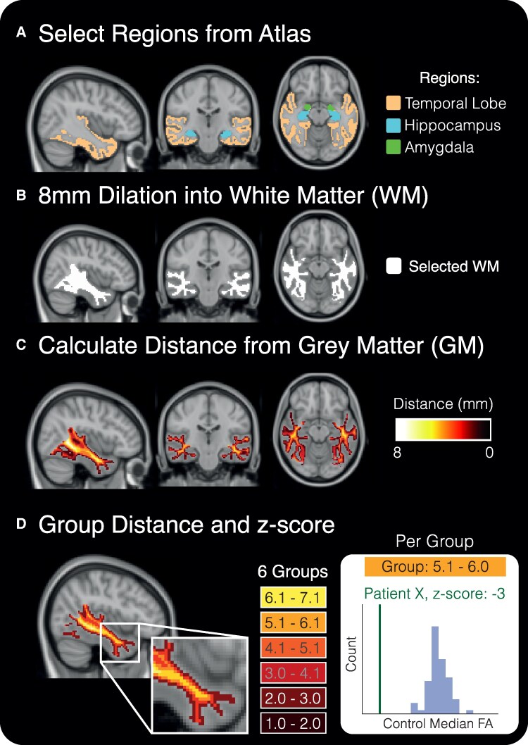
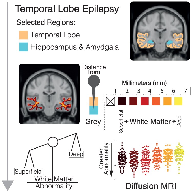
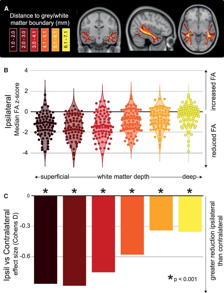
Non-invasive neuroimaging is important in epilepsy to help identify cerebral abnormalities. Abnormally reduced fractional anisotropy (FA) in deep white matter (WM) from diffusion-weighted imaging (DWI) is widely reported in large multi-cohort studies across all types of epilepsies. However, abnormalities in FA for superficial WM are rarely investigated in epilepsy. To gain a greater understanding of the nature of WM abnormality at different WM depths, we investigated DWI abnormalities at a range of superficial and deep WM in two separate temporal lobe epilepsy (TLE) cohorts. The first cohort (TLE = 81, Healthy Control; HC = 67) underwent a high angular resolution multi-shell DWI, whilst the second cohort (TLE = 70, HC = 29) had a single-shell acquisition. We registered FA maps to a standard template, and analysed temporal WM within 8 mm of the temporal lobe grey matter, amygdala and hippocampus. We standardised FA measures at different depths, and compared ipsi-versus contralateral temporal WM, and MRI-positive versus MRI-negative groups. We report three major findings: First, superficial WM had greater FA reductions than deep WM in TLE (P < 0.001). Second, this effect was more prominent in the ipsilateral than contralateral temporal lobe WM (P < 0.001). Third, these effects were present to a similar degree in patients who reported an MRI negative. All results are held in both TLE cohorts. These findings suggest that, in the temporal lobe, superficial WM is more abnormal than deep WM in TLE, with potential clinical use for lateralisation even in MRI-negative patients. These findings motivate further investigation of the importance of superficial WM in epilepsy.

 求助内容:
求助内容: 应助结果提醒方式:


