Recurrent Calcifying Pseudoneoplasm of the Neuraxis in Meckel's Cave with Cyst Extending into the Cerebellopontine Cistern after Resection of the Cystic Component: A Case Report.
{"title":"Recurrent Calcifying Pseudoneoplasm of the Neuraxis in Meckel's Cave with Cyst Extending into the Cerebellopontine Cistern after Resection of the Cystic Component: A Case Report.","authors":"Ryuta Yamada, Hiroki Kobayashi, Yuichi Nomura, Naoki Oka, Jouji Kokuzawa, Yasuhiko Kaku","doi":"10.1055/s-0045-1806859","DOIUrl":null,"url":null,"abstract":"<p><p>Calcifying pseudoneoplasms of the neuraxis (CAPNON) are rare non-neoplastic calcified lesions that can occur throughout the entire neuraxis. Cranial nerves may be involved in skull base lesions. Surgical resection usually has a good prognosis, with only a few cases of recurrence reported in the relevant literature. We experienced a recurrent case of CAPNON in Meckel's cave extending to the cerebellopontine cistern with involvement of the trigeminal nerve after initial resection of the cystic part of the lesion. A 48-year-old man presented with a 4-year history of right-sided ptosis. Magnetic resonance imaging revealed a calcified lesion arising in Meckel's cave, with a cystic lesion extending to the cerebellopontine cistern. The cystic part of the lesion was excised during the initial surgery using the right lateral suboccipital approach. However, the cystic lesion regrew to its preoperative size within 23 months. The right extradural subtemporal approach was used to successfully remove both the calcified mass in the right Meckel cave and the cystic lesion extending to the right cerebellopontine cistern during the resection of the lesion. The patient's neurological symptoms resolved postoperatively. CAPNON was identified during the histopathological examination. This report describes a rare case of CAPNON in Meckel's cave with a unique clinical course. After the first operation, cystic lesions recurred in the cerebellopontine cistern, but they were successfully treated by a second operation.</p>","PeriodicalId":94300,"journal":{"name":"Asian journal of neurosurgery","volume":"20 3","pages":"605-609"},"PeriodicalIF":0.0000,"publicationDate":"2025-03-25","publicationTypes":"Journal Article","fieldsOfStudy":null,"isOpenAccess":false,"openAccessPdf":"https://www.ncbi.nlm.nih.gov/pmc/articles/PMC12370357/pdf/","citationCount":"0","resultStr":null,"platform":"Semanticscholar","paperid":null,"PeriodicalName":"Asian journal of neurosurgery","FirstCategoryId":"1085","ListUrlMain":"https://doi.org/10.1055/s-0045-1806859","RegionNum":0,"RegionCategory":null,"ArticlePicture":[],"TitleCN":null,"AbstractTextCN":null,"PMCID":null,"EPubDate":"2025/9/1 0:00:00","PubModel":"eCollection","JCR":"","JCRName":"","Score":null,"Total":0}
引用次数: 0
Abstract
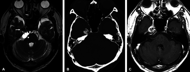
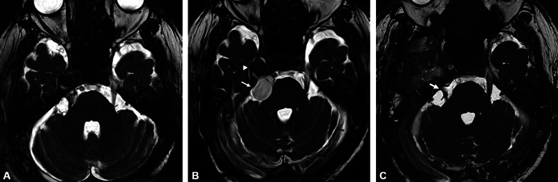
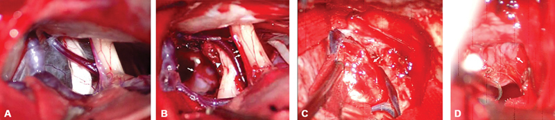
Calcifying pseudoneoplasms of the neuraxis (CAPNON) are rare non-neoplastic calcified lesions that can occur throughout the entire neuraxis. Cranial nerves may be involved in skull base lesions. Surgical resection usually has a good prognosis, with only a few cases of recurrence reported in the relevant literature. We experienced a recurrent case of CAPNON in Meckel's cave extending to the cerebellopontine cistern with involvement of the trigeminal nerve after initial resection of the cystic part of the lesion. A 48-year-old man presented with a 4-year history of right-sided ptosis. Magnetic resonance imaging revealed a calcified lesion arising in Meckel's cave, with a cystic lesion extending to the cerebellopontine cistern. The cystic part of the lesion was excised during the initial surgery using the right lateral suboccipital approach. However, the cystic lesion regrew to its preoperative size within 23 months. The right extradural subtemporal approach was used to successfully remove both the calcified mass in the right Meckel cave and the cystic lesion extending to the right cerebellopontine cistern during the resection of the lesion. The patient's neurological symptoms resolved postoperatively. CAPNON was identified during the histopathological examination. This report describes a rare case of CAPNON in Meckel's cave with a unique clinical course. After the first operation, cystic lesions recurred in the cerebellopontine cistern, but they were successfully treated by a second operation.

 求助内容:
求助内容: 应助结果提醒方式:


