Probable cerebral amyloid angiopathy-related inflammation presenting with dissociated hemorrhagic and inflammatory lesions in a middle-aged patient: A case report.
{"title":"Probable cerebral amyloid angiopathy-related inflammation presenting with dissociated hemorrhagic and inflammatory lesions in a middle-aged patient: A case report.","authors":"Toshihide Takahashi, Kiyoyuki Yanaka, Hitoshi Aiyama, Minami Saura, Hayato Takeda, Nobuyuki Takahashi, Aiki Marushima, Eiichi Ishikawa","doi":"10.25259/SNI_589_2025","DOIUrl":null,"url":null,"abstract":"<p><strong>Background: </strong>Cerebral amyloid angiopathy-related inflammation (CAA-ri) is a rare inflammatory encephalopathy associated with amyloid-β deposition in cerebral vessels, typically presenting in older adults with subacute cognitive decline, seizures, and lobar hemorrhages. It affects approximately 5% of patients with CAA, often showing asymmetric white matter hyperintensities, microbleeds, and leptomeningeal enhancement on magnetic resonance imaging (MRI). Vascular fragility in patients with CAA-ri increases rebleeding risk compared with those without CAA-ri, such as those with hypertensive intracerebral hemorrhage. This report describes a rare probable CAA-ri case in a middle-aged patient with an atypical presentation: younger age, absence of typical inflammatory symptoms, and spatially dissociated hemorrhagic and inflammatory lesions.</p><p><strong>Case description: </strong>A 50-year-old man with a history of childhood right frontal hemorrhage and epilepsy presented with right upper-limb numbness. Computed tomography revealed a left parietal subcortical hemorrhage, and MRI showed bilateral occipital white matter hyperintensities, microbleeds, and right occipital leptomeningeal enhancement. Biopsy of the right occipital lesion confirmed amyloid-β deposition with mild perivascular lymphocytic infiltration, indicating probable CAA-ri. He was managed conservatively owing to the minimal mass effect and received corticosteroids as outpatients. MRI at 19 months revealed resolution of occipital hyperintensities, without neurological deficits.</p><p><strong>Conclusion: </strong>This case highlights probable CAA-ri in a middle-aged adult, presenting with subcortical hemorrhage and spatially dissociated inflammatory lesions. Despite the subtle inflammatory findings, clinical, imaging, and histological evaluations supported the diagnosis. Early recognition and immunosuppressive therapy can achieve favorable outcomes, emphasizing CAA-ri's inclusion in the differential diagnosis of lobar hemorrhage in relatively younger patients without hypertension or vascular risk factors.</p>","PeriodicalId":94217,"journal":{"name":"Surgical neurology international","volume":"16 ","pages":"302"},"PeriodicalIF":0.0000,"publicationDate":"2025-07-25","publicationTypes":"Journal Article","fieldsOfStudy":null,"isOpenAccess":false,"openAccessPdf":"https://www.ncbi.nlm.nih.gov/pmc/articles/PMC12361660/pdf/","citationCount":"0","resultStr":null,"platform":"Semanticscholar","paperid":null,"PeriodicalName":"Surgical neurology international","FirstCategoryId":"1085","ListUrlMain":"https://doi.org/10.25259/SNI_589_2025","RegionNum":0,"RegionCategory":null,"ArticlePicture":[],"TitleCN":null,"AbstractTextCN":null,"PMCID":null,"EPubDate":"2025/1/1 0:00:00","PubModel":"eCollection","JCR":"","JCRName":"","Score":null,"Total":0}
引用次数: 0
Abstract
Background: Cerebral amyloid angiopathy-related inflammation (CAA-ri) is a rare inflammatory encephalopathy associated with amyloid-β deposition in cerebral vessels, typically presenting in older adults with subacute cognitive decline, seizures, and lobar hemorrhages. It affects approximately 5% of patients with CAA, often showing asymmetric white matter hyperintensities, microbleeds, and leptomeningeal enhancement on magnetic resonance imaging (MRI). Vascular fragility in patients with CAA-ri increases rebleeding risk compared with those without CAA-ri, such as those with hypertensive intracerebral hemorrhage. This report describes a rare probable CAA-ri case in a middle-aged patient with an atypical presentation: younger age, absence of typical inflammatory symptoms, and spatially dissociated hemorrhagic and inflammatory lesions.
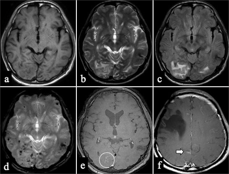
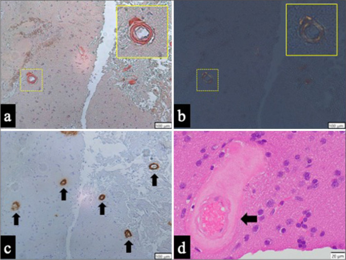
Case description: A 50-year-old man with a history of childhood right frontal hemorrhage and epilepsy presented with right upper-limb numbness. Computed tomography revealed a left parietal subcortical hemorrhage, and MRI showed bilateral occipital white matter hyperintensities, microbleeds, and right occipital leptomeningeal enhancement. Biopsy of the right occipital lesion confirmed amyloid-β deposition with mild perivascular lymphocytic infiltration, indicating probable CAA-ri. He was managed conservatively owing to the minimal mass effect and received corticosteroids as outpatients. MRI at 19 months revealed resolution of occipital hyperintensities, without neurological deficits.
Conclusion: This case highlights probable CAA-ri in a middle-aged adult, presenting with subcortical hemorrhage and spatially dissociated inflammatory lesions. Despite the subtle inflammatory findings, clinical, imaging, and histological evaluations supported the diagnosis. Early recognition and immunosuppressive therapy can achieve favorable outcomes, emphasizing CAA-ri's inclusion in the differential diagnosis of lobar hemorrhage in relatively younger patients without hypertension or vascular risk factors.


 求助内容:
求助内容: 应助结果提醒方式:


