{"title":"Intrinsic brainstem epidermoid cyst in childhood.","authors":"Tybault Hollanders, Sarah Hendrickx, Edward Baert","doi":"10.25259/SNI_198_2025","DOIUrl":null,"url":null,"abstract":"<p><strong>Background: </strong>Epidermoid cysts are slow-growing, rare congenital lesions. They are most seen in the cerebellopontine angle, fourth ventricle, or parasellar regions. Patients typically become symptomatic between 20- and 40 years of age. We present a rare case of a symptomatic intrinsic brainstem epidermoid cyst in a 12-year-old girl with atypical radiological features.</p><p><strong>Case description: </strong>A 12-year-old girl presented with progressive gait disturbances, disequilibrium, diplopia due to right-sided abducens paresis, left-sided facial paresis (HB grade II), headaches, nausea, and vomiting over the past 6 months. Magnetic resonance imaging (MRI) revealed an intrinsic lesion of the brainstem not present on MRI 4 years prior. A microsurgical gross total resection, including resection of cyst wall, was performed. The pathology report diagnosed the lesion as an epidermoid cyst. The patient improved significantly after resection.</p><p><strong>Conclusion: </strong>This pathology represents <1% of all intracranial tumors, and with only 21 documented pediatric intrinsic brainstem cases in medical literature, very rarely has an intrinsic brainstem location. We detail the medical history, work-up, surgical management, and postoperative outcomes, contributing to the limited body of knowledge regarding this exceptional entity.</p>","PeriodicalId":94217,"journal":{"name":"Surgical neurology international","volume":"16 ","pages":"290"},"PeriodicalIF":0.0000,"publicationDate":"2025-07-18","publicationTypes":"Journal Article","fieldsOfStudy":null,"isOpenAccess":false,"openAccessPdf":"https://www.ncbi.nlm.nih.gov/pmc/articles/PMC12361696/pdf/","citationCount":"0","resultStr":null,"platform":"Semanticscholar","paperid":null,"PeriodicalName":"Surgical neurology international","FirstCategoryId":"1085","ListUrlMain":"https://doi.org/10.25259/SNI_198_2025","RegionNum":0,"RegionCategory":null,"ArticlePicture":[],"TitleCN":null,"AbstractTextCN":null,"PMCID":null,"EPubDate":"2025/1/1 0:00:00","PubModel":"eCollection","JCR":"","JCRName":"","Score":null,"Total":0}
引用次数: 0
Abstract
Background: Epidermoid cysts are slow-growing, rare congenital lesions. They are most seen in the cerebellopontine angle, fourth ventricle, or parasellar regions. Patients typically become symptomatic between 20- and 40 years of age. We present a rare case of a symptomatic intrinsic brainstem epidermoid cyst in a 12-year-old girl with atypical radiological features.
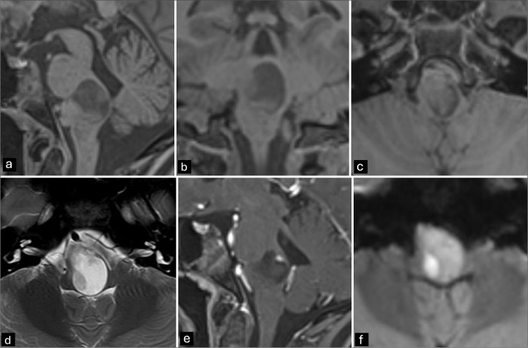
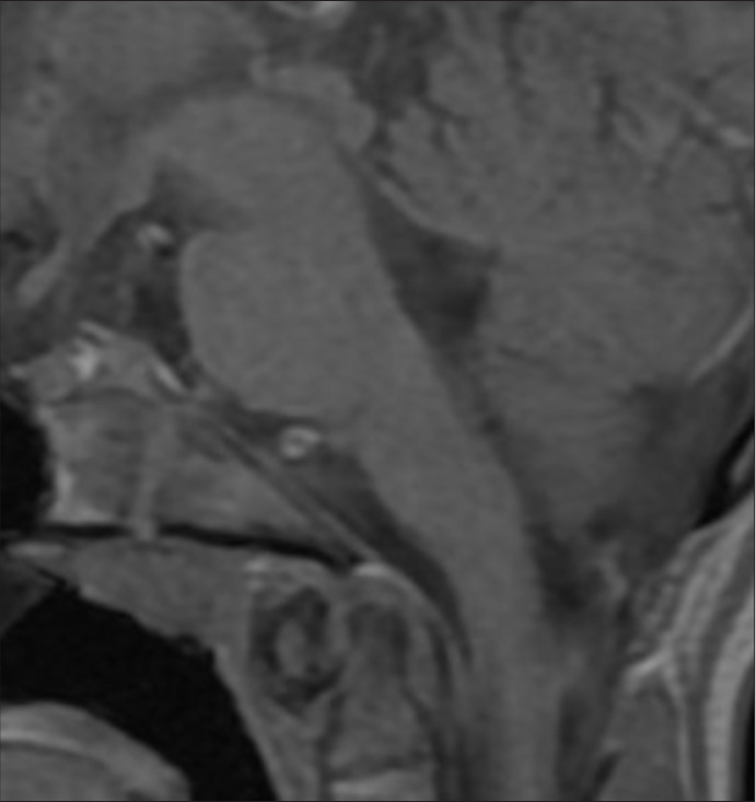
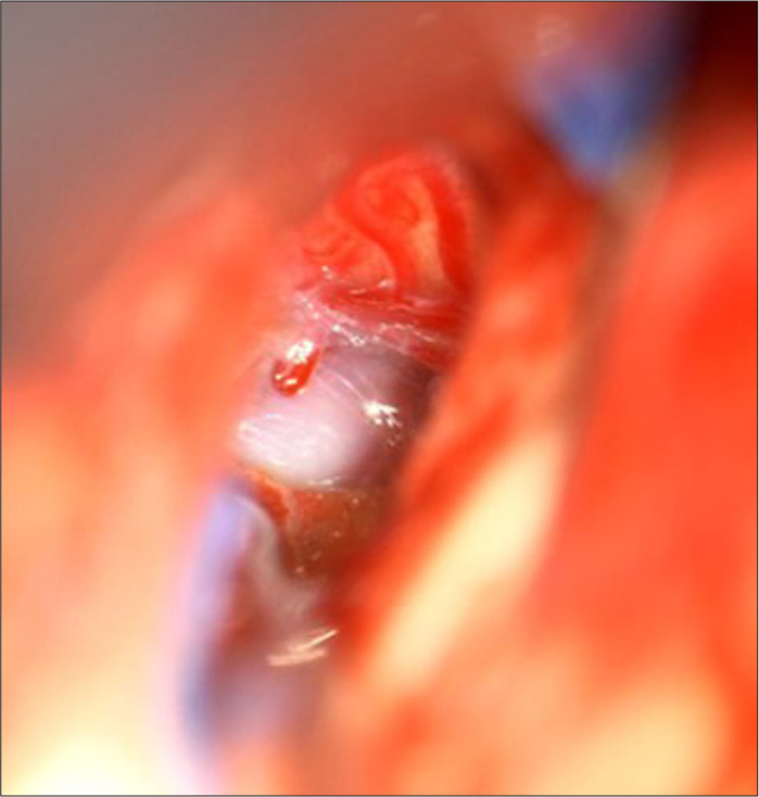
Case description: A 12-year-old girl presented with progressive gait disturbances, disequilibrium, diplopia due to right-sided abducens paresis, left-sided facial paresis (HB grade II), headaches, nausea, and vomiting over the past 6 months. Magnetic resonance imaging (MRI) revealed an intrinsic lesion of the brainstem not present on MRI 4 years prior. A microsurgical gross total resection, including resection of cyst wall, was performed. The pathology report diagnosed the lesion as an epidermoid cyst. The patient improved significantly after resection.
Conclusion: This pathology represents <1% of all intracranial tumors, and with only 21 documented pediatric intrinsic brainstem cases in medical literature, very rarely has an intrinsic brainstem location. We detail the medical history, work-up, surgical management, and postoperative outcomes, contributing to the limited body of knowledge regarding this exceptional entity.

 求助内容:
求助内容: 应助结果提醒方式:


