A Comprehensive Case Report of Metastatic Intracranial Melanoma with Brief Review of Literature.
Asian journal of neurosurgery
Pub Date : 2025-06-04
eCollection Date: 2025-09-01
DOI:10.1055/s-0045-1809329
引用次数: 0
Abstract
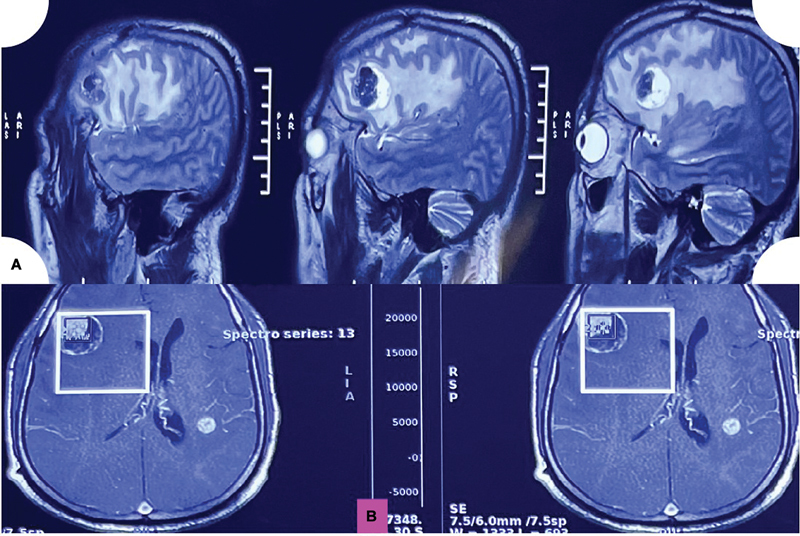
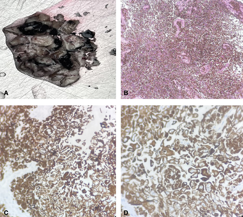
Primary intracranial melanomas are an extremely rare entity and are a diagnosis of exclusion. Malignant melanoma represents the third most common site for cerebral metastasis. We hereby narrate a comprehensive and detailed case of metastatic intracranial melanomas with BRAF mutation, which later on had an extensive systemic spread. The imaging differentials include metastasis, intracranial hemorrhage, or granuloma. The final and definitive diagnosis was attained by detailed clinical, histological, and immunohistochemical evaluation as metastatic malignant pigmented tumor consistent with intracranial melanoma.
转移性颅内黑色素瘤1例综合报道并文献复习。
原发性颅内黑色素瘤是一种极为罕见的疾病,诊断时需排除。恶性黑色素瘤是大脑转移的第三大常见部位。我们在此叙述一个全面而详细的病例转移性颅内黑色素瘤与BRAF突变,后来有广泛的全身扩散。影像学鉴别包括转移、颅内出血或肉芽肿。通过详细的临床、组织学和免疫组织化学评估,最终确诊为转移性恶性色素瘤,与颅内黑色素瘤一致。
本文章由计算机程序翻译,如有差异,请以英文原文为准。
求助全文
约1分钟内获得全文
求助全文

 求助内容:
求助内容: 应助结果提醒方式:


