Rahi Navelkar, Andrea Cosolo, Bogdan Bintu, Yubao Cheng, Vincent Gardeux, Silvia Gutnik, Taihei Fujimori, Antonina Hafner, Atishay Jay, Bojing Blair Jia, Adam Paul Jussila, Gerard Llimos, Antonios Lioutas, Nuno M C Martins, William J Moore, Yodai Takei, Frances Wong, Kaifu Yang, Huaiying Zhang, Quan Zhu, Magda Bienko, Lacramioara Bintu, Long Cai, Bart Deplancke, Marcelo Nollmann, Susan E Mango, Bing Ren, Peter J Park, Ahilya N Sawh, Andrew Schroeder, Jason R Swedlow, Golnaz Vahedi, Chao-Ting Wu, Sarah Aufmkolk, Alistair N Boettiger, Irene Farabella, Caterina Strambio-De-Castillia, Siyuan Wang
{"title":"FAIR sharing of Chromatin Tracing datasets using the newly developed 4DN FISH Omics Format.","authors":"Rahi Navelkar, Andrea Cosolo, Bogdan Bintu, Yubao Cheng, Vincent Gardeux, Silvia Gutnik, Taihei Fujimori, Antonina Hafner, Atishay Jay, Bojing Blair Jia, Adam Paul Jussila, Gerard Llimos, Antonios Lioutas, Nuno M C Martins, William J Moore, Yodai Takei, Frances Wong, Kaifu Yang, Huaiying Zhang, Quan Zhu, Magda Bienko, Lacramioara Bintu, Long Cai, Bart Deplancke, Marcelo Nollmann, Susan E Mango, Bing Ren, Peter J Park, Ahilya N Sawh, Andrew Schroeder, Jason R Swedlow, Golnaz Vahedi, Chao-Ting Wu, Sarah Aufmkolk, Alistair N Boettiger, Irene Farabella, Caterina Strambio-De-Castillia, Siyuan Wang","doi":"","DOIUrl":null,"url":null,"abstract":"<p><p>A key output of the NIH-Common Fund 4D Nucleome (4DN) project <sup>1,2</sup> is the open publication of datasets related to the structure of the human cell nucleus and the genome. Recent years have seen a rapid expansion of multiplexed Fluorescence In Situ Hybridization (FISH) or FISH-omics methods, which quantify the spatial organization of chromatin in single cells, sometimes together with RNA and protein measurements, and provide an expanded understanding of how 3D higher-order chromosome structure relates to transcriptional activity and cell development in both health and disease. Despite this progress, results from Chromatin Tracing FISH-omics experiments are difficult to share, reuse, and subject to third-party downstream analysis due to the lack of standard specifications for data exchange. Following up on the recent publication of Microscopy Metadata specifications <sup>3,4</sup>, we present the 4DN FISH Omics Format - Chromatin Tracing (FOF-CT), a community-developed data standard for processed results derived from a wide variety of imaging techniques for Chromatin Tracing, with the most recent studies falling roughly into two categories: ball-and-stick and volumetric based on whether they represent the targeted genomic segment as individual fluorescence spots or as clouds of single-molecule localizations. To demonstrate the value and potential use of FOF-CT to promote the FAIR sharing of the results obtained from Chromatin Tracing techniques, this manuscript will focus on ball-and-stick Chromatin Tracing techniques, including those described by the pioneering Chromatin Tracing study of Wang et al. <sup>5</sup> as well as Optical Reconstruction of Chromatin Architecture (ORCA) <sup>6</sup>, microscopy-based chromosome conformation capture (Hi-M) <sup>7</sup>, Multiplexed Imaging of Nucleome Architectures (MINA) <sup>8</sup>, DNA Sequential Fluorescence In Situ Hybridization (DNA seqFISH/seqFISH+) <sup>9-11</sup>, Oligopaint Fluorescent In Situ Sequencing (OligoFISSEQ) <sup>12</sup>, DNA Multiplexed error-robust fluorescence in situ hybridization (DNA-MERFISH) <sup>13</sup>, and In-situ Genomic Sequencing (IGS) <sup>14</sup>. The manuscript will describe the structure of the format and present a collection of FOF-CT datasets that were recently deposited to the 4DN Data Portal <sup>15</sup> and the Open Microscopy Environment (OME) Image Data Resource (IDR) platform <sup>16</sup> and are ideally suited for promoting reuse, exchange, further processing, and integrative modeling. Furthermore, the manuscript will present examples of analysis pipelines that could be applied more widely due to the existence of the FOF-CT exchange data format and provide examples of biological conclusions that could be drawn thanks to the availability of such datasets.</p>","PeriodicalId":93888,"journal":{"name":"ArXiv","volume":" ","pages":""},"PeriodicalIF":0.0000,"publicationDate":"2025-08-21","publicationTypes":"Journal Article","fieldsOfStudy":null,"isOpenAccess":false,"openAccessPdf":"https://www.ncbi.nlm.nih.gov/pmc/articles/PMC12393234/pdf/","citationCount":"0","resultStr":null,"platform":"Semanticscholar","paperid":null,"PeriodicalName":"ArXiv","FirstCategoryId":"1085","ListUrlMain":"","RegionNum":0,"RegionCategory":null,"ArticlePicture":[],"TitleCN":null,"AbstractTextCN":null,"PMCID":null,"EPubDate":"","PubModel":"","JCR":"","JCRName":"","Score":null,"Total":0}
引用次数: 0
Abstract
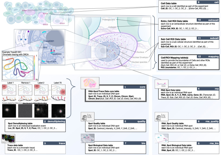
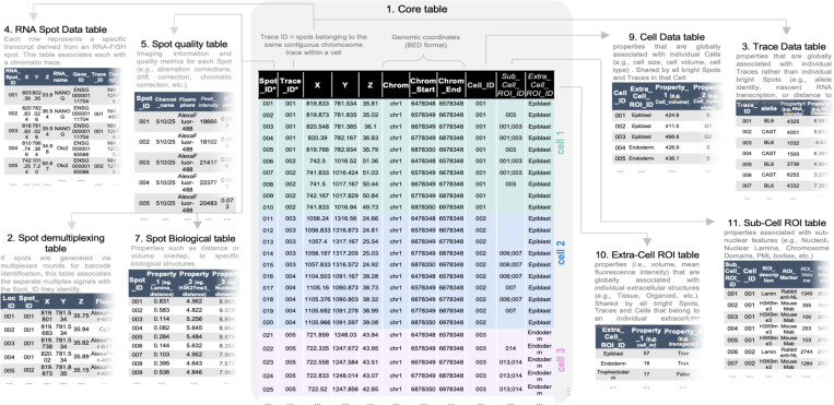
A key output of the NIH-Common Fund 4D Nucleome (4DN) project 1,2 is the open publication of datasets related to the structure of the human cell nucleus and the genome. Recent years have seen a rapid expansion of multiplexed Fluorescence In Situ Hybridization (FISH) or FISH-omics methods, which quantify the spatial organization of chromatin in single cells, sometimes together with RNA and protein measurements, and provide an expanded understanding of how 3D higher-order chromosome structure relates to transcriptional activity and cell development in both health and disease. Despite this progress, results from Chromatin Tracing FISH-omics experiments are difficult to share, reuse, and subject to third-party downstream analysis due to the lack of standard specifications for data exchange. Following up on the recent publication of Microscopy Metadata specifications 3,4, we present the 4DN FISH Omics Format - Chromatin Tracing (FOF-CT), a community-developed data standard for processed results derived from a wide variety of imaging techniques for Chromatin Tracing, with the most recent studies falling roughly into two categories: ball-and-stick and volumetric based on whether they represent the targeted genomic segment as individual fluorescence spots or as clouds of single-molecule localizations. To demonstrate the value and potential use of FOF-CT to promote the FAIR sharing of the results obtained from Chromatin Tracing techniques, this manuscript will focus on ball-and-stick Chromatin Tracing techniques, including those described by the pioneering Chromatin Tracing study of Wang et al. 5 as well as Optical Reconstruction of Chromatin Architecture (ORCA) 6, microscopy-based chromosome conformation capture (Hi-M) 7, Multiplexed Imaging of Nucleome Architectures (MINA) 8, DNA Sequential Fluorescence In Situ Hybridization (DNA seqFISH/seqFISH+) 9-11, Oligopaint Fluorescent In Situ Sequencing (OligoFISSEQ) 12, DNA Multiplexed error-robust fluorescence in situ hybridization (DNA-MERFISH) 13, and In-situ Genomic Sequencing (IGS) 14. The manuscript will describe the structure of the format and present a collection of FOF-CT datasets that were recently deposited to the 4DN Data Portal 15 and the Open Microscopy Environment (OME) Image Data Resource (IDR) platform 16 and are ideally suited for promoting reuse, exchange, further processing, and integrative modeling. Furthermore, the manuscript will present examples of analysis pipelines that could be applied more widely due to the existence of the FOF-CT exchange data format and provide examples of biological conclusions that could be drawn thanks to the availability of such datasets.

 求助内容:
求助内容: 应助结果提醒方式:


