{"title":"Diagnostic and clinical value of multiparameter magnetic resonance imaging in cesarean scar pregnancy: a comparative study of sequence combinations.","authors":"Xin-Lin Yao, Chun-Rong Wei, Yao-Yun Zhang, Si-Le Yin, Jian-Bo Wu, Jian-Yi Huang, Hong-Tao Liu, Mei-Ning Chen","doi":"10.21037/qims-2024-2589","DOIUrl":null,"url":null,"abstract":"<p><strong>Background: </strong>Cesarean scar pregnancy (CSP) is a special form of ectopic pregnancy that lacks specific clinical manifestations. Artificially induced abortion may lead to severe complications such as massive bleeding and even uterine rupture, posing a threat to the safety of pregnant women. Magnetic resonance imaging (MRI) has potential advantages in evaluating CSP. This study aimed to analyze the value of MRI with different combinations of sequences in diagnosing CSP following a cesarean section and to evaluate the clinical value of MRI in classifying CSP.</p><p><strong>Methods: </strong>We conducted a retrospective analysis on the clinical and imaging data of 80 patients with suspected CSP on ultrasound examination. The MRI data of all patients were divided into four combinations: combination A, T1-weighted imaging (T1WI) + T2-weighted imaging (T2WI); combination B, T1WI + T2WI + diffusion-weighted imaging (DWI); combination C, T1WI + T2WI + dynamic contrast-enhanced (DCE) MRI; and combination D, T2WI + DWI + DCE-MRI. The differences between these MRI sequence combinations were compared. Imaging features were observed, recorded, and used for MRI classification. Differences in imaging features between MRI classifications were also compared to determine their clinical significance.</p><p><strong>Results: </strong>Of the 80 cases confirmed by postoperative pathology, 67 (83.75%) were CSP. The area under the curve for the combinations C and D was larger (0.966 and 0.979, respectively) than that for combination A (0.883). The sensitivity, specificity, positive predictive value, and negative predictive value for combinations C and D were higher (combination C: sensitivity 98.51%, specificity 92.31%, positive predictive value 98.53%, and negative predictive value 92.31%; combination D: sensitivity 95.52%, specificity 92.31%, positive predictive value 98.46%, and negative predictive value 80.00%). The distribution of CSP type I (filled type), type II (partially filled type), and type III (covered type) was 19.40%, 59.70%, and 20.90%, respectively. There was no statistically significant difference in the length of the contact surface between the gestational sac and the scar among the MRI-type groups (H =0.012; P=0.994). However, the minimum thickness of the scar at the implantation site of type I was less than that in type II (H =-16.192; P=0.028), and the area of the gestational sac in the sagittal position was smaller in type I than in type III (H =-24.467; P=0.003).</p><p><strong>Conclusions: </strong>The preferred MRI sequence combination for diagnosing CSP should be T2WI + DWI + DCE-MRI. MRI can effectively visualize the relationship between the gestational sac and the incisional diverticulum in CSP and facilitate imaging-based staging.</p>","PeriodicalId":54267,"journal":{"name":"Quantitative Imaging in Medicine and Surgery","volume":"15 9","pages":"8282-8291"},"PeriodicalIF":2.3000,"publicationDate":"2025-09-01","publicationTypes":"Journal Article","fieldsOfStudy":null,"isOpenAccess":false,"openAccessPdf":"https://www.ncbi.nlm.nih.gov/pmc/articles/PMC12397649/pdf/","citationCount":"0","resultStr":null,"platform":"Semanticscholar","paperid":null,"PeriodicalName":"Quantitative Imaging in Medicine and Surgery","FirstCategoryId":"3","ListUrlMain":"https://doi.org/10.21037/qims-2024-2589","RegionNum":2,"RegionCategory":"医学","ArticlePicture":[],"TitleCN":null,"AbstractTextCN":null,"PMCID":null,"EPubDate":"2025/7/28 0:00:00","PubModel":"Epub","JCR":"Q2","JCRName":"RADIOLOGY, NUCLEAR MEDICINE & MEDICAL IMAGING","Score":null,"Total":0}
引用次数: 0
Abstract
Background: Cesarean scar pregnancy (CSP) is a special form of ectopic pregnancy that lacks specific clinical manifestations. Artificially induced abortion may lead to severe complications such as massive bleeding and even uterine rupture, posing a threat to the safety of pregnant women. Magnetic resonance imaging (MRI) has potential advantages in evaluating CSP. This study aimed to analyze the value of MRI with different combinations of sequences in diagnosing CSP following a cesarean section and to evaluate the clinical value of MRI in classifying CSP.
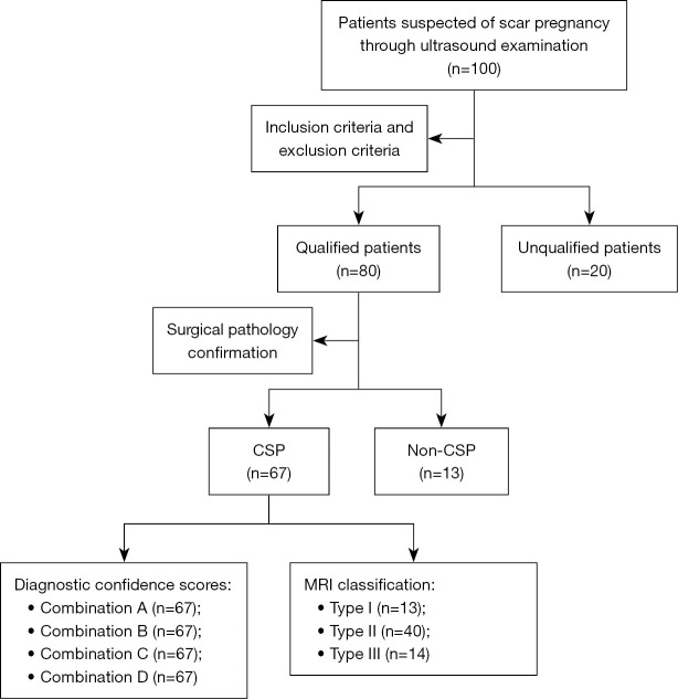
Methods: We conducted a retrospective analysis on the clinical and imaging data of 80 patients with suspected CSP on ultrasound examination. The MRI data of all patients were divided into four combinations: combination A, T1-weighted imaging (T1WI) + T2-weighted imaging (T2WI); combination B, T1WI + T2WI + diffusion-weighted imaging (DWI); combination C, T1WI + T2WI + dynamic contrast-enhanced (DCE) MRI; and combination D, T2WI + DWI + DCE-MRI. The differences between these MRI sequence combinations were compared. Imaging features were observed, recorded, and used for MRI classification. Differences in imaging features between MRI classifications were also compared to determine their clinical significance.
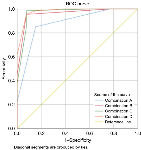
Results: Of the 80 cases confirmed by postoperative pathology, 67 (83.75%) were CSP. The area under the curve for the combinations C and D was larger (0.966 and 0.979, respectively) than that for combination A (0.883). The sensitivity, specificity, positive predictive value, and negative predictive value for combinations C and D were higher (combination C: sensitivity 98.51%, specificity 92.31%, positive predictive value 98.53%, and negative predictive value 92.31%; combination D: sensitivity 95.52%, specificity 92.31%, positive predictive value 98.46%, and negative predictive value 80.00%). The distribution of CSP type I (filled type), type II (partially filled type), and type III (covered type) was 19.40%, 59.70%, and 20.90%, respectively. There was no statistically significant difference in the length of the contact surface between the gestational sac and the scar among the MRI-type groups (H =0.012; P=0.994). However, the minimum thickness of the scar at the implantation site of type I was less than that in type II (H =-16.192; P=0.028), and the area of the gestational sac in the sagittal position was smaller in type I than in type III (H =-24.467; P=0.003).
Conclusions: The preferred MRI sequence combination for diagnosing CSP should be T2WI + DWI + DCE-MRI. MRI can effectively visualize the relationship between the gestational sac and the incisional diverticulum in CSP and facilitate imaging-based staging.

 求助内容:
求助内容: 应助结果提醒方式:


