Development of a prediction model for HER2 low breast cancer using quantitative intra- and peri-tumoral heterogeneity and MRI features on high-spatial resolution ultrafast DCE-MRI.
IF 2.3 2区 医学Q2 RADIOLOGY, NUCLEAR MEDICINE & MEDICAL IMAGING
Hongbing Luo, Shixuan Zhao, Zhe Chen, Juan Ji, Jing Ren, Yongjie Li, Peng Zhou
{"title":"Development of a prediction model for HER2 low breast cancer using quantitative intra- and peri-tumoral heterogeneity and MRI features on high-spatial resolution ultrafast DCE-MRI.","authors":"Hongbing Luo, Shixuan Zhao, Zhe Chen, Juan Ji, Jing Ren, Yongjie Li, Peng Zhou","doi":"10.21037/qims-24-976","DOIUrl":null,"url":null,"abstract":"<p><strong>Background: </strong>Accurate preoperative human epidermal growth factor receptor 2 (HER2) status assessment is crucial for guiding treatment selection, particularly with the emergence of anti-HER2 antibody-drug conjugates (ADCs) for HER2-low breast cancer. However, current immunohistochemistry (IHC)-based classification is limited by spatial heterogeneity and sampling bias. Quantitative analysis of intra- and peri-tumoral heterogeneity (ITH) on imaging may offer a non-invasive, objective, and reproducible approach to distinguish HER2-low breast cancer from other subtypes. This study aimed to investigate quantitative ITH from high-spatial resolution ultrafast dynamic contrast-enhanced magnetic resonance imaging (UF DCE-MRI) based kinetic curves in distinguishing HER2 low from HER2 zero or positive breast cancer.</p><p><strong>Methods: </strong>Consecutive breast cancer patients who underwent preoperative high-spatial-resolution UF DCE-MRI were retrospectively enrolled. They were stratified into HER2 zero, HER2 low, or HER2 positive groups based on IHC and in situ hybridization results. Traditional MRI findings and clinicopathological characteristics were evaluated, and personalized ITH scores were constructed using semi-quantitative parameters derived from kinetic curves. Models incorporating ITH, MRI, and clinicopathological distinctions were developed for dichotomized HER2 statuses prediction using multivariable logistic regression. The added value of ITH in the Final Combined Model was evaluated.</p><p><strong>Results: </strong>This study enrolled 368 patients, with 45.9% (169/368) having HER2-low breast cancer. The ITH score was higher in HER2 low than that in HER2 zero (P<0.001), but lower than that in HER2 positive (P<0.001). The ITH score was higher in HER2 positive compared to HER2 zero (P<0.001). The Final Combined Model integrating ITH, MRI, and clinicopathological variables achieved good predictive performance, achieving area under the curve (AUC) values of 0.80 [95% confidence interval (CI): 0.75-0.86] for HER2 low <i>vs.</i> zero, 0.85 (95% CI: 0.80-0.89) for HER2 low <i>vs.</i> positive, and 0.83 (95% CI: 0.77-0.88) for HER2 zero <i>vs.</i> positive. The corresponding sensitivity/specificity values were 77%/72%, 77%/81%, and 94%/58%, respectively. The ITH score significantly enhanced HER2 status prediction, supported by AUC improvement (DeLong test, P<0.05), along with statistical significance in net reclassification improvement (NRI) (P<0.001) and integrated discrimination improvement (IDI) (P<0.001) across all tasks.</p><p><strong>Conclusions: </strong>Integrating ITH from high-spatial resolution UF DCE-MRI-based kinetic curves improved the non-invasive differentiation of HER2-low breast cancer. This approach may guide targeted biopsy strategies and aid in selecting candidates for anti-HER2 ADC therapy, optimizing HER2-targeted precision medicine.</p>","PeriodicalId":54267,"journal":{"name":"Quantitative Imaging in Medicine and Surgery","volume":"15 9","pages":"7788-7802"},"PeriodicalIF":2.3000,"publicationDate":"2025-09-01","publicationTypes":"Journal Article","fieldsOfStudy":null,"isOpenAccess":false,"openAccessPdf":"https://www.ncbi.nlm.nih.gov/pmc/articles/PMC12397656/pdf/","citationCount":"0","resultStr":null,"platform":"Semanticscholar","paperid":null,"PeriodicalName":"Quantitative Imaging in Medicine and Surgery","FirstCategoryId":"3","ListUrlMain":"https://doi.org/10.21037/qims-24-976","RegionNum":2,"RegionCategory":"医学","ArticlePicture":[],"TitleCN":null,"AbstractTextCN":null,"PMCID":null,"EPubDate":"2025/8/18 0:00:00","PubModel":"Epub","JCR":"Q2","JCRName":"RADIOLOGY, NUCLEAR MEDICINE & MEDICAL IMAGING","Score":null,"Total":0}
引用次数: 0
Abstract
Background: Accurate preoperative human epidermal growth factor receptor 2 (HER2) status assessment is crucial for guiding treatment selection, particularly with the emergence of anti-HER2 antibody-drug conjugates (ADCs) for HER2-low breast cancer. However, current immunohistochemistry (IHC)-based classification is limited by spatial heterogeneity and sampling bias. Quantitative analysis of intra- and peri-tumoral heterogeneity (ITH) on imaging may offer a non-invasive, objective, and reproducible approach to distinguish HER2-low breast cancer from other subtypes. This study aimed to investigate quantitative ITH from high-spatial resolution ultrafast dynamic contrast-enhanced magnetic resonance imaging (UF DCE-MRI) based kinetic curves in distinguishing HER2 low from HER2 zero or positive breast cancer.
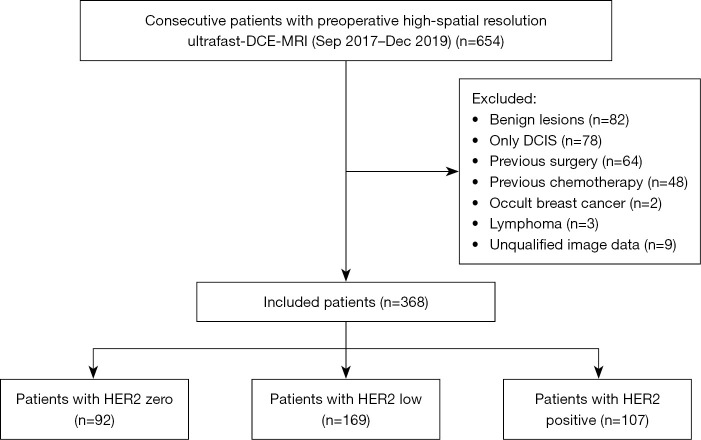
Methods: Consecutive breast cancer patients who underwent preoperative high-spatial-resolution UF DCE-MRI were retrospectively enrolled. They were stratified into HER2 zero, HER2 low, or HER2 positive groups based on IHC and in situ hybridization results. Traditional MRI findings and clinicopathological characteristics were evaluated, and personalized ITH scores were constructed using semi-quantitative parameters derived from kinetic curves. Models incorporating ITH, MRI, and clinicopathological distinctions were developed for dichotomized HER2 statuses prediction using multivariable logistic regression. The added value of ITH in the Final Combined Model was evaluated.
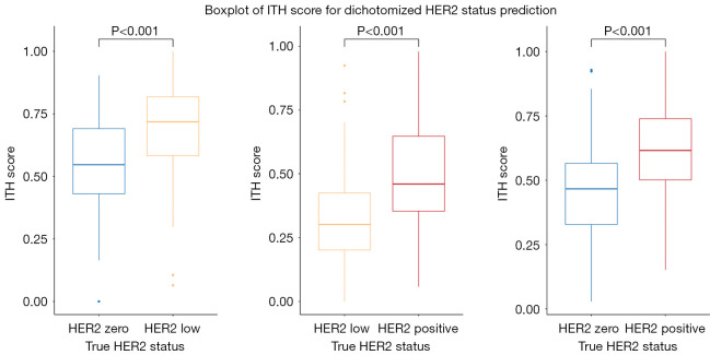
Results: This study enrolled 368 patients, with 45.9% (169/368) having HER2-low breast cancer. The ITH score was higher in HER2 low than that in HER2 zero (P<0.001), but lower than that in HER2 positive (P<0.001). The ITH score was higher in HER2 positive compared to HER2 zero (P<0.001). The Final Combined Model integrating ITH, MRI, and clinicopathological variables achieved good predictive performance, achieving area under the curve (AUC) values of 0.80 [95% confidence interval (CI): 0.75-0.86] for HER2 low vs. zero, 0.85 (95% CI: 0.80-0.89) for HER2 low vs. positive, and 0.83 (95% CI: 0.77-0.88) for HER2 zero vs. positive. The corresponding sensitivity/specificity values were 77%/72%, 77%/81%, and 94%/58%, respectively. The ITH score significantly enhanced HER2 status prediction, supported by AUC improvement (DeLong test, P<0.05), along with statistical significance in net reclassification improvement (NRI) (P<0.001) and integrated discrimination improvement (IDI) (P<0.001) across all tasks.
Conclusions: Integrating ITH from high-spatial resolution UF DCE-MRI-based kinetic curves improved the non-invasive differentiation of HER2-low breast cancer. This approach may guide targeted biopsy strategies and aid in selecting candidates for anti-HER2 ADC therapy, optimizing HER2-targeted precision medicine.

 求助内容:
求助内容: 应助结果提醒方式:


