Enhancing postoperative recurrence assessment in gastric and colorectal cancer patients with intraperitoneal fluorouracil implants: overcoming the diagnostic challenge of fluorouracil implant-related tumor-like lesions.
IF 2.3 2区 医学Q2 RADIOLOGY, NUCLEAR MEDICINE & MEDICAL IMAGING
{"title":"Enhancing postoperative recurrence assessment in gastric and colorectal cancer patients with intraperitoneal fluorouracil implants: overcoming the diagnostic challenge of fluorouracil implant-related tumor-like lesions.","authors":"Luwen Hao, Xin Chen, Sijia Zhou, Xuemei Hu, Daoyu Hu, Zhen Li, Yaqi Shen","doi":"10.21037/qims-24-2033","DOIUrl":null,"url":null,"abstract":"<p><strong>Background: </strong>Intraoperative intraperitoneal chemotherapy using sustained-release fluorouracil implants has been used to reduce the recurrence of gastrointestinal tumors. However, these implants may persist and present as tumor-like lesions in imaging studies, potentially leading to false-positive interpretations as metastatic sites, affecting patient management. Our study aimed to enhance the diagnostic accuracy of radiologists in assessing gastric and colorectal cancer patients with fluorouracil implants.</p><p><strong>Methods: </strong>This retrospective study comprised a summary of fluorouracil implant-related lesion characteristics by a multidisciplinary team (MDT), and a three-stage evaluation of tumor-like lesions by two radiologists. In total, 240 computed tomography (CT) examinations were randomly selected from all the available CT examinations of all patients, whom were then further divided evenly into three groups. Two radiologists independently assessed the implant-related tumor-like lesions across the following three stages: stage 1: pre-training without surgical information; stage 2: post-training without surgical information; and stage 3: post-training with surgical details provided. The training was based on the characteristics of the lesions identified earlier by the MDT. The radiologists evaluated the malignancy or benignity of each lesion, and rated their diagnostic confidence using a three-point scale. The reference standard was determined by the MDT. Diagnostic accuracy and diagnostic confidence were compared using Pearson's Chi-squared test and the Wilcoxon rank-sum test.</p><p><strong>Results: </strong>A total of 168 fluorouracil implants were confirmed in the subdiaphragmatic regions, paracolic gutters, and tumor beds of 164 patients. Imaging features such as a typical foreign body reaction (85.71%), no contrast enhancement on CT/magnetic resonance imaging, and no diffusion restriction on diffusion-weighted imaging were important for differentiating between fluorouracil implant-related lesions and malignant lesions. Follow-up CT scans showed a size reduction in 67.26% of the lesions and density changes in 52.98%. The diagnostic accuracy and confidence of the radiologists were improved in stage 2 (accuracy: 91.25%; confidence: most often classified as medium) compared to stage 1 (accuracy: 67.5%; confidence: most often classified as low; both P<0.001). When surgical information was available, the diagnostic accuracy and confidence of the radiologists were improved in stage 3 (accuracy: 100%; confidence most often classified as high) compared to stage 2 (accuracy: P=0.007; confidence: P<0.001).</p><p><strong>Conclusions: </strong>The diagnostic accuracy and confidence of radiologists can be improved by providing them with training on implant imaging characteristics and precise surgical record documentation on the implant location and quantity.</p>","PeriodicalId":54267,"journal":{"name":"Quantitative Imaging in Medicine and Surgery","volume":"15 9","pages":"7774-7787"},"PeriodicalIF":2.3000,"publicationDate":"2025-09-01","publicationTypes":"Journal Article","fieldsOfStudy":null,"isOpenAccess":false,"openAccessPdf":"https://www.ncbi.nlm.nih.gov/pmc/articles/PMC12397638/pdf/","citationCount":"0","resultStr":null,"platform":"Semanticscholar","paperid":null,"PeriodicalName":"Quantitative Imaging in Medicine and Surgery","FirstCategoryId":"3","ListUrlMain":"https://doi.org/10.21037/qims-24-2033","RegionNum":2,"RegionCategory":"医学","ArticlePicture":[],"TitleCN":null,"AbstractTextCN":null,"PMCID":null,"EPubDate":"2025/8/19 0:00:00","PubModel":"Epub","JCR":"Q2","JCRName":"RADIOLOGY, NUCLEAR MEDICINE & MEDICAL IMAGING","Score":null,"Total":0}
引用次数: 0
Abstract
Background: Intraoperative intraperitoneal chemotherapy using sustained-release fluorouracil implants has been used to reduce the recurrence of gastrointestinal tumors. However, these implants may persist and present as tumor-like lesions in imaging studies, potentially leading to false-positive interpretations as metastatic sites, affecting patient management. Our study aimed to enhance the diagnostic accuracy of radiologists in assessing gastric and colorectal cancer patients with fluorouracil implants.
Methods: This retrospective study comprised a summary of fluorouracil implant-related lesion characteristics by a multidisciplinary team (MDT), and a three-stage evaluation of tumor-like lesions by two radiologists. In total, 240 computed tomography (CT) examinations were randomly selected from all the available CT examinations of all patients, whom were then further divided evenly into three groups. Two radiologists independently assessed the implant-related tumor-like lesions across the following three stages: stage 1: pre-training without surgical information; stage 2: post-training without surgical information; and stage 3: post-training with surgical details provided. The training was based on the characteristics of the lesions identified earlier by the MDT. The radiologists evaluated the malignancy or benignity of each lesion, and rated their diagnostic confidence using a three-point scale. The reference standard was determined by the MDT. Diagnostic accuracy and diagnostic confidence were compared using Pearson's Chi-squared test and the Wilcoxon rank-sum test.
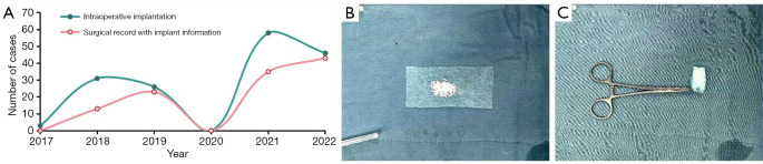
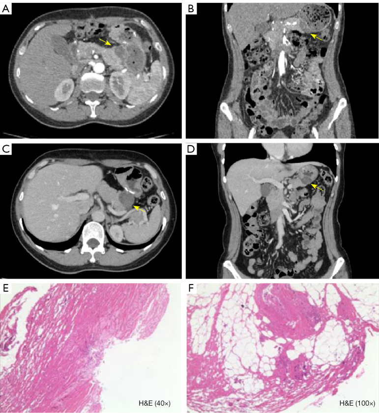
Results: A total of 168 fluorouracil implants were confirmed in the subdiaphragmatic regions, paracolic gutters, and tumor beds of 164 patients. Imaging features such as a typical foreign body reaction (85.71%), no contrast enhancement on CT/magnetic resonance imaging, and no diffusion restriction on diffusion-weighted imaging were important for differentiating between fluorouracil implant-related lesions and malignant lesions. Follow-up CT scans showed a size reduction in 67.26% of the lesions and density changes in 52.98%. The diagnostic accuracy and confidence of the radiologists were improved in stage 2 (accuracy: 91.25%; confidence: most often classified as medium) compared to stage 1 (accuracy: 67.5%; confidence: most often classified as low; both P<0.001). When surgical information was available, the diagnostic accuracy and confidence of the radiologists were improved in stage 3 (accuracy: 100%; confidence most often classified as high) compared to stage 2 (accuracy: P=0.007; confidence: P<0.001).
Conclusions: The diagnostic accuracy and confidence of radiologists can be improved by providing them with training on implant imaging characteristics and precise surgical record documentation on the implant location and quantity.

 求助内容:
求助内容: 应助结果提醒方式:


