Extracellular volume fraction based on dual-layer spectral computed tomography in assessment of gastric cancer: feasibility analysis of low-dose equilibrium phase scanning.
IF 2.3 2区 医学Q2 RADIOLOGY, NUCLEAR MEDICINE & MEDICAL IMAGING
{"title":"Extracellular volume fraction based on dual-layer spectral computed tomography in assessment of gastric cancer: feasibility analysis of low-dose equilibrium phase scanning.","authors":"Zhen Zhang, Xiaoping Zhao, Xuelian Chen, Hongyan Wang, Xiaohui Zhang, Yonggang Li, Mengzhe Zuo","doi":"10.21037/qims-24-2013","DOIUrl":null,"url":null,"abstract":"<p><strong>Background: </strong>The extracellular volume fraction (fECV) based on equilibrium phase iodine density images (IDIs) of dual-layer spectral detector computed tomography (DLCT) can be used in the assessment of gastric cancer (GC). However, obtaining the equilibrium phase images requires a higher radiation dose. The purpose of our study was to evaluate the feasibility of low-dose equilibrium phase scans on DLCT for fECV acquisition in histological grading assessment of GC.</p><p><strong>Methods: </strong>A total of 86 gastric adenocarcinoma patients confirmed by surgical pathology were divided into two groups that underwent contrast-enhanced DLCT with routine-dose (120 kV/129 refmAs) and low-dose (120 kV/90 refmAs) equilibrium phases, respectively. The fECV values of GC lesions were measured from IDIs in the equilibrium phase. The radiation dose, image quality of the equilibrium phase images, and fECV values were compared between the low- and routine-dose groups. Then, the performance of the fECV in the two groups to distinguish histological grades of GC lesions was evaluated using a receiver operating characteristic (ROC) curve and the DeLong test. The fECV maps were reconstructed from the IDIs of the equilibrium phase.</p><p><strong>Results: </strong>The radiation dose of the equilibrium phase and the accumulated dose in the low-dose group decreased by 54% and 34%, respectively, compared to the routine-dose group (both P<0.001). The image noise of equilibrium phase images was higher in the low-dose group than that in the routine-dose group (P<0.001) and the noise scores of the low-dose group were lower than those of the routine-dose group (P=0.003), whereas no significant differences were detected in the signal-to-noise ratio (SNR), contrast-to-noise ratio (CNR), detail score, and fECV values between the two groups (P=0.243, 0.607, 0.861, and 0.301, respectively). The fECV values of high-grade GC lesions were higher than those of the low-grade lesions in the two groups (52.98%±8.06% <i>vs.</i> 38.31%±5.24%, P<0.001, and 51.94%±9.11% <i>vs.</i> 36.91%±5.26%, P=0.002). The fECV obtained in the low-dose group had a similar performance compared to the routine-dose group in histological grading assessment of GC [area under the curve (AUC): 0.871 <i>vs.</i> 0.879, <i>Z</i>=-0.148, P=0.882].</p><p><strong>Conclusions: </strong>Contrast-enhanced DLCT with low-dose equilibrium phase scans in GC reduced the radiation dose while providing comparable image quality and performance of fECV in histological grading assessment to those of routine-dose scans.</p>","PeriodicalId":54267,"journal":{"name":"Quantitative Imaging in Medicine and Surgery","volume":"15 9","pages":"8529-8540"},"PeriodicalIF":2.3000,"publicationDate":"2025-09-01","publicationTypes":"Journal Article","fieldsOfStudy":null,"isOpenAccess":false,"openAccessPdf":"https://www.ncbi.nlm.nih.gov/pmc/articles/PMC12397705/pdf/","citationCount":"0","resultStr":null,"platform":"Semanticscholar","paperid":null,"PeriodicalName":"Quantitative Imaging in Medicine and Surgery","FirstCategoryId":"3","ListUrlMain":"https://doi.org/10.21037/qims-24-2013","RegionNum":2,"RegionCategory":"医学","ArticlePicture":[],"TitleCN":null,"AbstractTextCN":null,"PMCID":null,"EPubDate":"2025/8/15 0:00:00","PubModel":"Epub","JCR":"Q2","JCRName":"RADIOLOGY, NUCLEAR MEDICINE & MEDICAL IMAGING","Score":null,"Total":0}
引用次数: 0
Abstract
Background: The extracellular volume fraction (fECV) based on equilibrium phase iodine density images (IDIs) of dual-layer spectral detector computed tomography (DLCT) can be used in the assessment of gastric cancer (GC). However, obtaining the equilibrium phase images requires a higher radiation dose. The purpose of our study was to evaluate the feasibility of low-dose equilibrium phase scans on DLCT for fECV acquisition in histological grading assessment of GC.
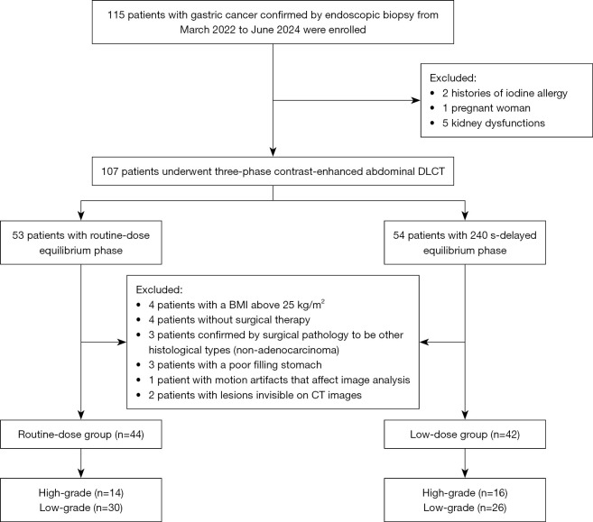
Methods: A total of 86 gastric adenocarcinoma patients confirmed by surgical pathology were divided into two groups that underwent contrast-enhanced DLCT with routine-dose (120 kV/129 refmAs) and low-dose (120 kV/90 refmAs) equilibrium phases, respectively. The fECV values of GC lesions were measured from IDIs in the equilibrium phase. The radiation dose, image quality of the equilibrium phase images, and fECV values were compared between the low- and routine-dose groups. Then, the performance of the fECV in the two groups to distinguish histological grades of GC lesions was evaluated using a receiver operating characteristic (ROC) curve and the DeLong test. The fECV maps were reconstructed from the IDIs of the equilibrium phase.
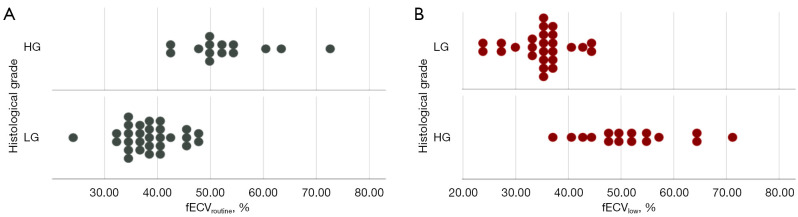
Results: The radiation dose of the equilibrium phase and the accumulated dose in the low-dose group decreased by 54% and 34%, respectively, compared to the routine-dose group (both P<0.001). The image noise of equilibrium phase images was higher in the low-dose group than that in the routine-dose group (P<0.001) and the noise scores of the low-dose group were lower than those of the routine-dose group (P=0.003), whereas no significant differences were detected in the signal-to-noise ratio (SNR), contrast-to-noise ratio (CNR), detail score, and fECV values between the two groups (P=0.243, 0.607, 0.861, and 0.301, respectively). The fECV values of high-grade GC lesions were higher than those of the low-grade lesions in the two groups (52.98%±8.06% vs. 38.31%±5.24%, P<0.001, and 51.94%±9.11% vs. 36.91%±5.26%, P=0.002). The fECV obtained in the low-dose group had a similar performance compared to the routine-dose group in histological grading assessment of GC [area under the curve (AUC): 0.871 vs. 0.879, Z=-0.148, P=0.882].
Conclusions: Contrast-enhanced DLCT with low-dose equilibrium phase scans in GC reduced the radiation dose while providing comparable image quality and performance of fECV in histological grading assessment to those of routine-dose scans.


 求助内容:
求助内容: 应助结果提醒方式:


