Evaluation of skeletal muscle microcirculation via magnetic resonance intravoxel incoherent motion perfusion imaging in an acute ischemia rabbit model.
IF 2.3 2区 医学Q2 RADIOLOGY, NUCLEAR MEDICINE & MEDICAL IMAGING
{"title":"Evaluation of skeletal muscle microcirculation via magnetic resonance intravoxel incoherent motion perfusion imaging in an acute ischemia rabbit model.","authors":"Guoping Song, Xinyu Song, Jienan Wang, Xiance Zhao, Junkang Shen","doi":"10.21037/qims-2025-711","DOIUrl":null,"url":null,"abstract":"<p><strong>Background: </strong>Acute limb ischemia (ALI) necessitates prompt intervention to prevent severe complications such as amputation. Current clinical assessments lack reliable quantitative methods for gauging skeletal muscle ischemia severity. Intravoxel incoherent motion (IVIM) perfusion imaging is a noninvasive approach for quantifying microvascular perfusion. We aimed to assess microcirculation alterations in rabbit vastus lateralis muscle following acute ischemia using IVIM.</p><p><strong>Methods: </strong>Acute ischemia models were established in 25 New Zealand white rabbits through arterial ligation of their right hind legs. Magnetic resonance imaging (MRI) examinations of the vastus lateralis muscle were conducted hourly postsurgery for a duration of 7 hours. The scan sequences included IVIM, adenosine triphosphate (APT), T1-weighted imaging (T1WI), T2-weighted imaging with fat suppression (T2WI-FS), T2 mapping, and diffusion-weighted imaging (DWI). The correlations between MRI results, ischemic time, and pathological changes were analyzed.</p><p><strong>Results: </strong>The perfusion fraction (f) of ischemic muscle significantly decreased from 6.19%±1.13% at 1 hour to 2.50%±0.64% at 7 hours (control: 7.19%±1.03%), representing a strong negative correlation with ischemic duration (r=-0.790). The true diffusion coefficient (D) remained relatively stable [(1.41-1.46)×10<sup>-3</sup> mm<sup>2</sup>/s] but was slightly elevated compared to controls. The pseudo-diffusion coefficient (D*) showed a sharp increase at 5 hours [(74.01±5.79)×10<sup>-3</sup> mm<sup>2</sup>/s]; control: [(61.28±9.31)×10<sup>-3</sup> mm<sup>2</sup>/s], followed by a drop at 6 hours [(59.44±15.77)×10<sup>-3</sup> mm<sup>2</sup>/s], suggesting sudden structural changes, which were confirmed by histopathology. T2WI-FS and DWI showed increased signal intensity in ischemic muscle, with the T2 relaxation times being significantly elevated (P<0.001) and positively correlated with ischemic duration (r=0.807). Apparent diffusion coefficient (ADC) values also increased with time (r=0.623). The amide proton transfer effect was enhanced in ischemic skeletal muscle throughout the 2-7-hour post-ischemic period (ischemic: 2.26%±0.39% at the 7th hour <i>vs</i>. control: 1.77%±0.33%; P<0.05).</p><p><strong>Conclusions: </strong>MRI effectively visualizes and detects skeletal muscle ischemia, with IVIM-derived f values providing a quantitative measure of microcirculatory impairment. D* potentially serves as a biomarker for identifying irreversible muscle fiber damage and may be a valuable tool for the quantitative assessment of ischemic injury to skeletal muscle.</p>","PeriodicalId":54267,"journal":{"name":"Quantitative Imaging in Medicine and Surgery","volume":"15 9","pages":"8064-8078"},"PeriodicalIF":2.3000,"publicationDate":"2025-09-01","publicationTypes":"Journal Article","fieldsOfStudy":null,"isOpenAccess":false,"openAccessPdf":"https://www.ncbi.nlm.nih.gov/pmc/articles/PMC12397635/pdf/","citationCount":"0","resultStr":null,"platform":"Semanticscholar","paperid":null,"PeriodicalName":"Quantitative Imaging in Medicine and Surgery","FirstCategoryId":"3","ListUrlMain":"https://doi.org/10.21037/qims-2025-711","RegionNum":2,"RegionCategory":"医学","ArticlePicture":[],"TitleCN":null,"AbstractTextCN":null,"PMCID":null,"EPubDate":"2025/8/13 0:00:00","PubModel":"Epub","JCR":"Q2","JCRName":"RADIOLOGY, NUCLEAR MEDICINE & MEDICAL IMAGING","Score":null,"Total":0}
引用次数: 0
Abstract
Background: Acute limb ischemia (ALI) necessitates prompt intervention to prevent severe complications such as amputation. Current clinical assessments lack reliable quantitative methods for gauging skeletal muscle ischemia severity. Intravoxel incoherent motion (IVIM) perfusion imaging is a noninvasive approach for quantifying microvascular perfusion. We aimed to assess microcirculation alterations in rabbit vastus lateralis muscle following acute ischemia using IVIM.
Methods: Acute ischemia models were established in 25 New Zealand white rabbits through arterial ligation of their right hind legs. Magnetic resonance imaging (MRI) examinations of the vastus lateralis muscle were conducted hourly postsurgery for a duration of 7 hours. The scan sequences included IVIM, adenosine triphosphate (APT), T1-weighted imaging (T1WI), T2-weighted imaging with fat suppression (T2WI-FS), T2 mapping, and diffusion-weighted imaging (DWI). The correlations between MRI results, ischemic time, and pathological changes were analyzed.
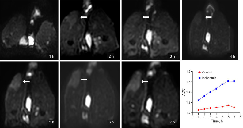
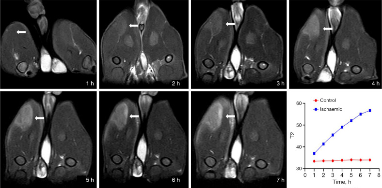
Results: The perfusion fraction (f) of ischemic muscle significantly decreased from 6.19%±1.13% at 1 hour to 2.50%±0.64% at 7 hours (control: 7.19%±1.03%), representing a strong negative correlation with ischemic duration (r=-0.790). The true diffusion coefficient (D) remained relatively stable [(1.41-1.46)×10-3 mm2/s] but was slightly elevated compared to controls. The pseudo-diffusion coefficient (D*) showed a sharp increase at 5 hours [(74.01±5.79)×10-3 mm2/s]; control: [(61.28±9.31)×10-3 mm2/s], followed by a drop at 6 hours [(59.44±15.77)×10-3 mm2/s], suggesting sudden structural changes, which were confirmed by histopathology. T2WI-FS and DWI showed increased signal intensity in ischemic muscle, with the T2 relaxation times being significantly elevated (P<0.001) and positively correlated with ischemic duration (r=0.807). Apparent diffusion coefficient (ADC) values also increased with time (r=0.623). The amide proton transfer effect was enhanced in ischemic skeletal muscle throughout the 2-7-hour post-ischemic period (ischemic: 2.26%±0.39% at the 7th hour vs. control: 1.77%±0.33%; P<0.05).
Conclusions: MRI effectively visualizes and detects skeletal muscle ischemia, with IVIM-derived f values providing a quantitative measure of microcirculatory impairment. D* potentially serves as a biomarker for identifying irreversible muscle fiber damage and may be a valuable tool for the quantitative assessment of ischemic injury to skeletal muscle.


 求助内容:
求助内容: 应助结果提醒方式:


