Comparing respiratory-triggered T2WI MRI with an artificial intelligence-assisted technique and motion-suppressed respiratory-triggered T2WI in abdominal imaging.
IF 2.3 2区 医学Q2 RADIOLOGY, NUCLEAR MEDICINE & MEDICAL IMAGING
Nan Wang, Yuhui Liu, Jiangnan Ran, Qi An, Lihua Chen, Ying Zhao, Dan Yu, Ailian Liu, Lina Zhuang, Qingwei Song
{"title":"Comparing respiratory-triggered T2WI MRI with an artificial intelligence-assisted technique and motion-suppressed respiratory-triggered T2WI in abdominal imaging.","authors":"Nan Wang, Yuhui Liu, Jiangnan Ran, Qi An, Lihua Chen, Ying Zhao, Dan Yu, Ailian Liu, Lina Zhuang, Qingwei Song","doi":"10.21037/qims-2025-71","DOIUrl":null,"url":null,"abstract":"<p><strong>Background: </strong>Magnetic resonance imaging (MRI) plays a crucial role in the diagnosis of abdominal conditions. A comprehensive assessment, especially of the liver, requires multi-planar T2-weighted sequences. To mitigate the effect of respiratory motion on image quality, the combination of acquisition and reconstruction with motion suppression (ARMS) and respiratory triggering (RT) is commonly employed. While this method maintains image quality, it does so at the expense of longer acquisition times. We evaluated the effectiveness of free-breathing, artificial intelligence-assisted compressed-sensing respiratory-triggered T2-weighted imaging (ACS-RT T2WI) compared to conventional acquisition and reconstruction with motion-suppression respiratory-triggered T2-weighted imaging (ARMS-RT T2WI) in abdominal MRI, assessing both qualitative and quantitative measures of image quality and lesion detection.</p><p><strong>Methods: </strong>In this retrospective study, 334 patients with upper abdominal discomfort were examined on a 3.0T MRI system. Each patient underwent both ARMS-RT T2WI and ACS-RT T2WI. Image quality was analyzed by two independent readers using a five-point Likert scale. The quantitative measurements included the signal-to-noise ratio (SNR), contrast-to-noise ratio (CNR), peak signal-to-noise ratio (PSNR), and sharpness. Lesion detection rates and contrast ratios (CRs) were also evaluated for liver, biliary system, and pancreatic lesions.</p><p><strong>Results: </strong>There ACS-RT T2WI protocol had a significantly reduced median scanning time compared to the ARMS-RT T2WI protocol (148.22±38.37 <i>vs.</i> 13.86±1.72 seconds). However, ARMS-RT T2WI had a higher PSNR than ACS-RT T2WI (39.87±2.72 <i>vs.</i> 38.69±3.00, P<0.05). Of the 201 liver lesions, ARMS-RT T2WI detected 193 (96.0%) and ACS-RT T2WI detected 192 (95.5%) (P=0.787). Of the 97 biliary system lesions, ARMS-RT T2WI detected 92 (94.8%) and ACS-RT T2WI detected 94 (96.9%) (P=0.721). Of the 110 pancreatic lesions, ARMS-RT T2WI detected 102 (92.7%) and ACS-RT T2WI detected 104 (94.5%) (P=0.784). The CR analysis showed the superior performance of ACS-RT T2WI in certain lesion types (hemangioma, 0.58±0.11 <i>vs.</i> 0.55±0.12; biliary tumor, 0.47±0.09 <i>vs.</i> 0.38±0.09; pancreatic cystic lesions, 0.59±0.12 <i>vs.</i> 0.48±0.14; pancreatic cancer, 0.48±0.18 <i>vs.</i> 0.43±0.17), but no significant difference was found in others like focal nodular hyperplasia (FNH), hepatapostema, hepatocellular carcinoma (HCC), cholangiocarcinoma, metastatic tumors, and biliary calculus.</p><p><strong>Conclusions: </strong>ACS-RT T2WI ensures clinical reliability with a substantial scan time reduction (>80%). Despite minor losses in detail and SNR reduction, ACS-RT T2WI does not impair lesion detection, marking its efficacy in abdominal imaging.</p>","PeriodicalId":54267,"journal":{"name":"Quantitative Imaging in Medicine and Surgery","volume":"15 9","pages":"7761-7773"},"PeriodicalIF":2.3000,"publicationDate":"2025-09-01","publicationTypes":"Journal Article","fieldsOfStudy":null,"isOpenAccess":false,"openAccessPdf":"https://www.ncbi.nlm.nih.gov/pmc/articles/PMC12397657/pdf/","citationCount":"0","resultStr":null,"platform":"Semanticscholar","paperid":null,"PeriodicalName":"Quantitative Imaging in Medicine and Surgery","FirstCategoryId":"3","ListUrlMain":"https://doi.org/10.21037/qims-2025-71","RegionNum":2,"RegionCategory":"医学","ArticlePicture":[],"TitleCN":null,"AbstractTextCN":null,"PMCID":null,"EPubDate":"2025/8/19 0:00:00","PubModel":"Epub","JCR":"Q2","JCRName":"RADIOLOGY, NUCLEAR MEDICINE & MEDICAL IMAGING","Score":null,"Total":0}
引用次数: 0
Abstract
Background: Magnetic resonance imaging (MRI) plays a crucial role in the diagnosis of abdominal conditions. A comprehensive assessment, especially of the liver, requires multi-planar T2-weighted sequences. To mitigate the effect of respiratory motion on image quality, the combination of acquisition and reconstruction with motion suppression (ARMS) and respiratory triggering (RT) is commonly employed. While this method maintains image quality, it does so at the expense of longer acquisition times. We evaluated the effectiveness of free-breathing, artificial intelligence-assisted compressed-sensing respiratory-triggered T2-weighted imaging (ACS-RT T2WI) compared to conventional acquisition and reconstruction with motion-suppression respiratory-triggered T2-weighted imaging (ARMS-RT T2WI) in abdominal MRI, assessing both qualitative and quantitative measures of image quality and lesion detection.
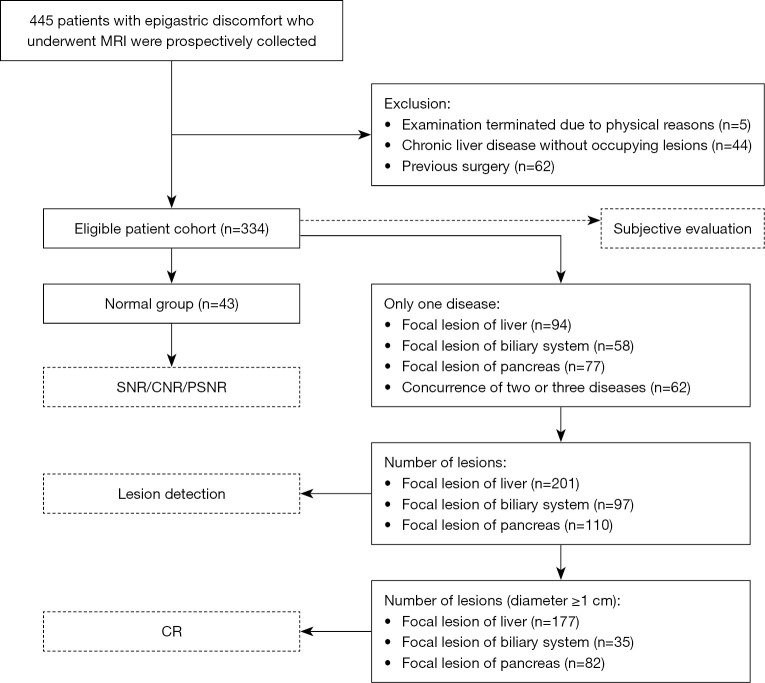
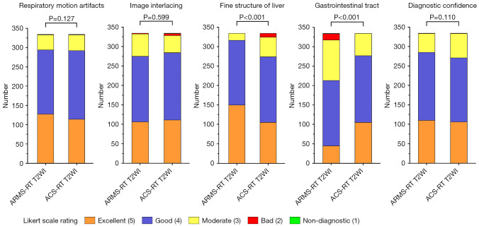
Methods: In this retrospective study, 334 patients with upper abdominal discomfort were examined on a 3.0T MRI system. Each patient underwent both ARMS-RT T2WI and ACS-RT T2WI. Image quality was analyzed by two independent readers using a five-point Likert scale. The quantitative measurements included the signal-to-noise ratio (SNR), contrast-to-noise ratio (CNR), peak signal-to-noise ratio (PSNR), and sharpness. Lesion detection rates and contrast ratios (CRs) were also evaluated for liver, biliary system, and pancreatic lesions.
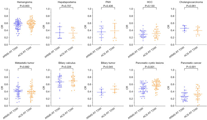
Results: There ACS-RT T2WI protocol had a significantly reduced median scanning time compared to the ARMS-RT T2WI protocol (148.22±38.37 vs. 13.86±1.72 seconds). However, ARMS-RT T2WI had a higher PSNR than ACS-RT T2WI (39.87±2.72 vs. 38.69±3.00, P<0.05). Of the 201 liver lesions, ARMS-RT T2WI detected 193 (96.0%) and ACS-RT T2WI detected 192 (95.5%) (P=0.787). Of the 97 biliary system lesions, ARMS-RT T2WI detected 92 (94.8%) and ACS-RT T2WI detected 94 (96.9%) (P=0.721). Of the 110 pancreatic lesions, ARMS-RT T2WI detected 102 (92.7%) and ACS-RT T2WI detected 104 (94.5%) (P=0.784). The CR analysis showed the superior performance of ACS-RT T2WI in certain lesion types (hemangioma, 0.58±0.11 vs. 0.55±0.12; biliary tumor, 0.47±0.09 vs. 0.38±0.09; pancreatic cystic lesions, 0.59±0.12 vs. 0.48±0.14; pancreatic cancer, 0.48±0.18 vs. 0.43±0.17), but no significant difference was found in others like focal nodular hyperplasia (FNH), hepatapostema, hepatocellular carcinoma (HCC), cholangiocarcinoma, metastatic tumors, and biliary calculus.
Conclusions: ACS-RT T2WI ensures clinical reliability with a substantial scan time reduction (>80%). Despite minor losses in detail and SNR reduction, ACS-RT T2WI does not impair lesion detection, marking its efficacy in abdominal imaging.

 求助内容:
求助内容: 应助结果提醒方式:


