An exploratory study of the diffusion tensor imaging analysis along perivascular spaces (DTI-ALPS) index combined with quantitative analysis of choroid plexus volume and perivascular spaces in different cognitive stages of cerebral small vessel disease.
IF 2.3 2区 医学Q2 RADIOLOGY, NUCLEAR MEDICINE & MEDICAL IMAGING
Wenli Lu, Li Yang, Ran Chen, Xinyi Chen, Shengnan Zhu, Liya Ji, Han Liao, Jing Qiang, Wenyi Li, Cheng Li, Dan Zhou
{"title":"An exploratory study of the diffusion tensor imaging analysis along perivascular spaces (DTI-ALPS) index combined with quantitative analysis of choroid plexus volume and perivascular spaces in different cognitive stages of cerebral small vessel disease.","authors":"Wenli Lu, Li Yang, Ran Chen, Xinyi Chen, Shengnan Zhu, Liya Ji, Han Liao, Jing Qiang, Wenyi Li, Cheng Li, Dan Zhou","doi":"10.21037/qims-2025-733","DOIUrl":null,"url":null,"abstract":"<p><strong>Background: </strong>Cerebral small vessel disease (CSVD) is a major contributor to cognitive impairment and dementia. Growing evidence suggests that impaired perivascular clearance plays a pivotal role in CSVD pathogenesis, yet non-invasive biomarkers for early cognitive decline remain limited. This study aimed to explore the diagnostic value of the diffusion tensor imaging analysis along perivascular spaces (DTI-ALPS) index, enlarged perivascular spaces (EPVS) numbers/volume, and choroid plexus volume (CPV) across different cognitive stages of CSVD.</p><p><strong>Methods: </strong>We retrospectively analyzed data from 102 CSVD patients [33 CSVD-cognitive normal (CSVD-CN); 39 CSVD-mild cognitive impairment (CSVD-MCI); 30 vascular dementia (VaD)] and 29 normal controls (NCs). Quantitative measurements of the DTI-ALPS index, EPVS numbers/volume, and CPV were obtained. Correlations with Montreal Cognitive Assessment (MoCA) scores and diagnostic performance were also evaluated.</p><p><strong>Results: </strong>Progressive DTI-ALPS index reduction (NCs: 1.50±0.19, CSVD-CN: 1.41±0.17, CSVD-MCI: 1.34±0.16, VaD: 1.33±0.17; P<0.001, r=0.36) and increases in basal ganglia (BG)-EPVS numbers {NCs: 4 [3, 6], CSVD-CN: 14 [10, 17], CSVD-MCI: 16 [12, 25], VaD: 22 [13, 31]; P<0.001, r=-0.45} and CPV {NCs: 1.35 [1.02, 1.65] cm<sup>3</sup>, CSVD-CN: 1.39 [1.11, 1.72] cm<sup>3</sup>, CSVD-MCI: 1.88 [1.41, 2.94] cm<sup>3</sup>, VaD: 2.89 [2.09, 3.39] cm<sup>3</sup>; P<0.001, r=-0.43} correlated with cognitive decline. BG-EPVS numbers excellently distinguished CSVD from NCs [area under the receiver operating characteristic (ROC) curve (AUC) =0.926; 95% confidence interval (CI): 0.882-0.971; sensitivity =84.2%; specificity =89.7%]. CPV emerged as the optimal standalone biomarker for VaD (AUC =0.758; 95% CI: 0.647-0.869; sensitivity =82.8%; specificity =69.5%). The multiparametric model (DTI-ALPS + BG-EPVS numbers + CPV) achieved high diagnostic accuracy: NCs <i>vs.</i> CSVD: AUC =0.978, 95% CI: 0.958-0.998; VaD <i>vs.</i> NCs/CSVD-CN/CSVD-MCI: AUC =0.825, 95% CI: 0.747-0.903; CSVD-MCI/VaD <i>vs.</i> NCs/CSVD-CN: AUC =0.900, 95% CI: 0.847-0.952.</p><p><strong>Conclusions: </strong>Combining the DTI-ALPS index, BG-EPVS numbers, and CPV may enhance early diagnosis and subtype differentiation in CSVD-related cognitive impairment, supporting targeted interventions.</p>","PeriodicalId":54267,"journal":{"name":"Quantitative Imaging in Medicine and Surgery","volume":"15 9","pages":"8173-8188"},"PeriodicalIF":2.3000,"publicationDate":"2025-09-01","publicationTypes":"Journal Article","fieldsOfStudy":null,"isOpenAccess":false,"openAccessPdf":"https://www.ncbi.nlm.nih.gov/pmc/articles/PMC12397676/pdf/","citationCount":"0","resultStr":null,"platform":"Semanticscholar","paperid":null,"PeriodicalName":"Quantitative Imaging in Medicine and Surgery","FirstCategoryId":"3","ListUrlMain":"https://doi.org/10.21037/qims-2025-733","RegionNum":2,"RegionCategory":"医学","ArticlePicture":[],"TitleCN":null,"AbstractTextCN":null,"PMCID":null,"EPubDate":"2025/8/15 0:00:00","PubModel":"Epub","JCR":"Q2","JCRName":"RADIOLOGY, NUCLEAR MEDICINE & MEDICAL IMAGING","Score":null,"Total":0}
引用次数: 0
Abstract
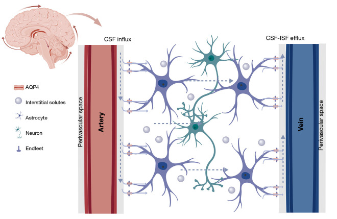
Background: Cerebral small vessel disease (CSVD) is a major contributor to cognitive impairment and dementia. Growing evidence suggests that impaired perivascular clearance plays a pivotal role in CSVD pathogenesis, yet non-invasive biomarkers for early cognitive decline remain limited. This study aimed to explore the diagnostic value of the diffusion tensor imaging analysis along perivascular spaces (DTI-ALPS) index, enlarged perivascular spaces (EPVS) numbers/volume, and choroid plexus volume (CPV) across different cognitive stages of CSVD.
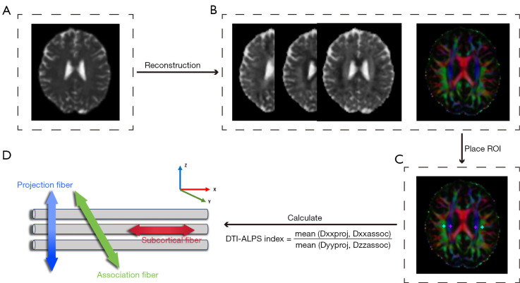
Methods: We retrospectively analyzed data from 102 CSVD patients [33 CSVD-cognitive normal (CSVD-CN); 39 CSVD-mild cognitive impairment (CSVD-MCI); 30 vascular dementia (VaD)] and 29 normal controls (NCs). Quantitative measurements of the DTI-ALPS index, EPVS numbers/volume, and CPV were obtained. Correlations with Montreal Cognitive Assessment (MoCA) scores and diagnostic performance were also evaluated.
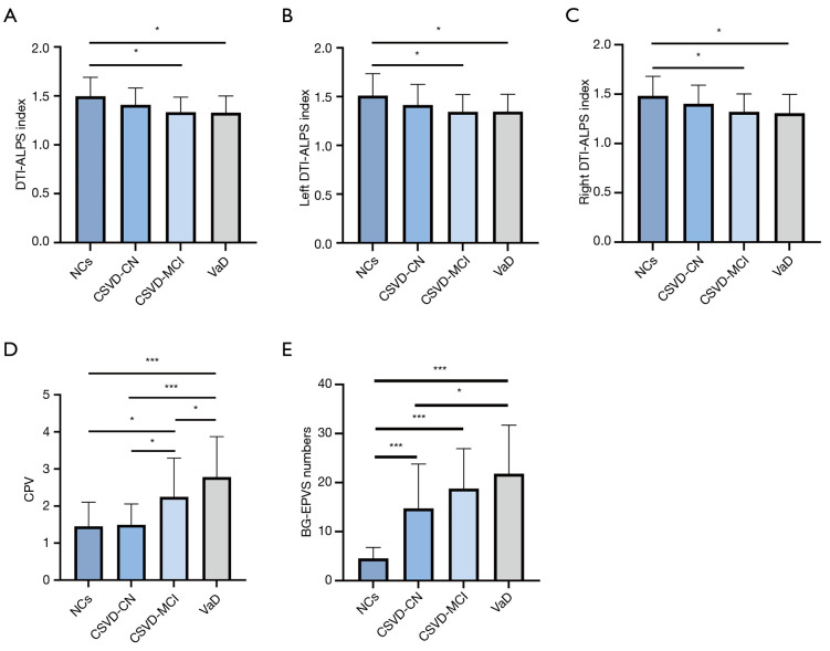
Results: Progressive DTI-ALPS index reduction (NCs: 1.50±0.19, CSVD-CN: 1.41±0.17, CSVD-MCI: 1.34±0.16, VaD: 1.33±0.17; P<0.001, r=0.36) and increases in basal ganglia (BG)-EPVS numbers {NCs: 4 [3, 6], CSVD-CN: 14 [10, 17], CSVD-MCI: 16 [12, 25], VaD: 22 [13, 31]; P<0.001, r=-0.45} and CPV {NCs: 1.35 [1.02, 1.65] cm3, CSVD-CN: 1.39 [1.11, 1.72] cm3, CSVD-MCI: 1.88 [1.41, 2.94] cm3, VaD: 2.89 [2.09, 3.39] cm3; P<0.001, r=-0.43} correlated with cognitive decline. BG-EPVS numbers excellently distinguished CSVD from NCs [area under the receiver operating characteristic (ROC) curve (AUC) =0.926; 95% confidence interval (CI): 0.882-0.971; sensitivity =84.2%; specificity =89.7%]. CPV emerged as the optimal standalone biomarker for VaD (AUC =0.758; 95% CI: 0.647-0.869; sensitivity =82.8%; specificity =69.5%). The multiparametric model (DTI-ALPS + BG-EPVS numbers + CPV) achieved high diagnostic accuracy: NCs vs. CSVD: AUC =0.978, 95% CI: 0.958-0.998; VaD vs. NCs/CSVD-CN/CSVD-MCI: AUC =0.825, 95% CI: 0.747-0.903; CSVD-MCI/VaD vs. NCs/CSVD-CN: AUC =0.900, 95% CI: 0.847-0.952.
Conclusions: Combining the DTI-ALPS index, BG-EPVS numbers, and CPV may enhance early diagnosis and subtype differentiation in CSVD-related cognitive impairment, supporting targeted interventions.

 求助内容:
求助内容: 应助结果提醒方式:


