{"title":"Disrupted neurovascular coupling in patients with lung cancer after chemotherapy.","authors":"Lanyue Hu, Shaohua Ding, Jun Yao, Yujie Zhang, Jia You, Huiyou Chen, Qian Li, Yu-Chen Chen, Xindao Yin","doi":"10.21037/qims-24-1321","DOIUrl":null,"url":null,"abstract":"<p><strong>Background: </strong>Chemotherapy-related cognitive impairments (CRCIs) are frequently reported by patients with non-small cell lung cancer (NSCLC) following chemotherapy treatment. Studies have revealed that cognitive impairment may be linked to abnormal spontaneous neuronal activity and changes in cerebral blood flow (CBF). However, the specific impact of neurovascular coupling (NVC) alterations on patients who have undergone chemotherapy has not been clarified. The aim of this study was to examine the variations in NVC in patients with lung cancer postchemotherapy and to determine potential correlations between these NVC alterations and neurocognitive dysfunction.</p><p><strong>Methods: </strong>A sample of 43 patients with NSCLC was recruited, including 20 patients treated with chemotherapy [CT(+)] and 23 chemotherapy-naïve [CT(-)] individuals who underwent pseudocontinuous arterial spin labeling (pCASL) scans and resting-state functional magnetic resonance imaging (rs-fMRI), along with neurocognitive evaluations. Global and regional NVC indices were assessed according to correlation coefficients and the ratios between CBF and neuronal activity-derived metrics, including the amplitude of low-frequency fluctuations (ALFF) and regional homogeneity (ReHo). Statistical analyses were conducted to calculate the difference between groups and characterize relationships between alterations in global and regional NVC and cognitive performance.</p><p><strong>Results: </strong>In comparison to the CT(-) group, the CT(+) group exhibited significantly lower coupling strength for global CBF-ALFF and CBF-ReHo correlations (P<0.05). Regionally, the CT(+) group demonstrated a decreased CBF:ALFF ratio in the right middle temporal gyrus (MTG) and left middle frontal gyrus (MFG), as well as an increased CBF:ALFF ratio in the left thalamus and left parahippocampal region. Furthermore, the CT(+) group had higher CBF:ReHo ratios in the left precuneus, right central operculum, right inferior parietal lobule, and right superior occipital gyrus but lower CBF:ReHo ratios in the left inferior frontal gyrus and right MFG (false-discovery rate-corrected P value <0.05). Notably, there was a negative correlation observed between Montreal Cognitive Assessment scores and memory scores and the CBF:ALFF ratios in the right MFG and left parahippocampal region.</p><p><strong>Conclusions: </strong>This research offers comprehensive insights into the neurological foundations of CRCI. The application of multimodal neuroimaging analyses combining rs-fMRI and pCASL may uncover the induction of neurovascular decoupling in lung cancer patients undergoing chemotherapy.</p>","PeriodicalId":54267,"journal":{"name":"Quantitative Imaging in Medicine and Surgery","volume":"15 9","pages":"7820-7832"},"PeriodicalIF":2.3000,"publicationDate":"2025-09-01","publicationTypes":"Journal Article","fieldsOfStudy":null,"isOpenAccess":false,"openAccessPdf":"https://www.ncbi.nlm.nih.gov/pmc/articles/PMC12397636/pdf/","citationCount":"0","resultStr":null,"platform":"Semanticscholar","paperid":null,"PeriodicalName":"Quantitative Imaging in Medicine and Surgery","FirstCategoryId":"3","ListUrlMain":"https://doi.org/10.21037/qims-24-1321","RegionNum":2,"RegionCategory":"医学","ArticlePicture":[],"TitleCN":null,"AbstractTextCN":null,"PMCID":null,"EPubDate":"2025/8/15 0:00:00","PubModel":"Epub","JCR":"Q2","JCRName":"RADIOLOGY, NUCLEAR MEDICINE & MEDICAL IMAGING","Score":null,"Total":0}
引用次数: 0
Abstract
Background: Chemotherapy-related cognitive impairments (CRCIs) are frequently reported by patients with non-small cell lung cancer (NSCLC) following chemotherapy treatment. Studies have revealed that cognitive impairment may be linked to abnormal spontaneous neuronal activity and changes in cerebral blood flow (CBF). However, the specific impact of neurovascular coupling (NVC) alterations on patients who have undergone chemotherapy has not been clarified. The aim of this study was to examine the variations in NVC in patients with lung cancer postchemotherapy and to determine potential correlations between these NVC alterations and neurocognitive dysfunction.
Methods: A sample of 43 patients with NSCLC was recruited, including 20 patients treated with chemotherapy [CT(+)] and 23 chemotherapy-naïve [CT(-)] individuals who underwent pseudocontinuous arterial spin labeling (pCASL) scans and resting-state functional magnetic resonance imaging (rs-fMRI), along with neurocognitive evaluations. Global and regional NVC indices were assessed according to correlation coefficients and the ratios between CBF and neuronal activity-derived metrics, including the amplitude of low-frequency fluctuations (ALFF) and regional homogeneity (ReHo). Statistical analyses were conducted to calculate the difference between groups and characterize relationships between alterations in global and regional NVC and cognitive performance.
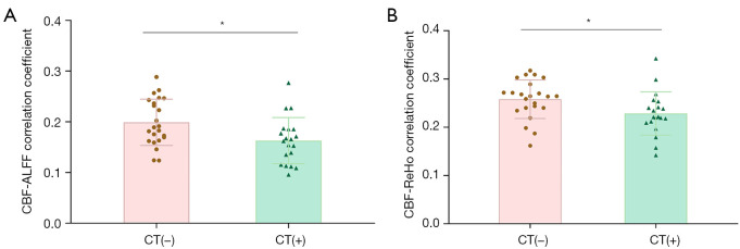
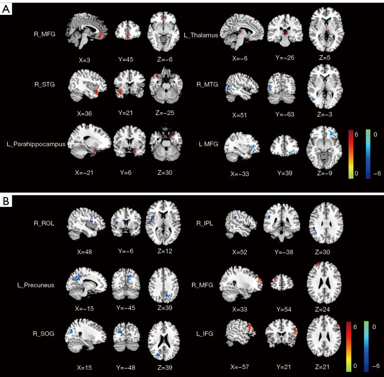
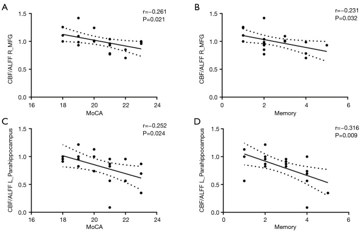
Results: In comparison to the CT(-) group, the CT(+) group exhibited significantly lower coupling strength for global CBF-ALFF and CBF-ReHo correlations (P<0.05). Regionally, the CT(+) group demonstrated a decreased CBF:ALFF ratio in the right middle temporal gyrus (MTG) and left middle frontal gyrus (MFG), as well as an increased CBF:ALFF ratio in the left thalamus and left parahippocampal region. Furthermore, the CT(+) group had higher CBF:ReHo ratios in the left precuneus, right central operculum, right inferior parietal lobule, and right superior occipital gyrus but lower CBF:ReHo ratios in the left inferior frontal gyrus and right MFG (false-discovery rate-corrected P value <0.05). Notably, there was a negative correlation observed between Montreal Cognitive Assessment scores and memory scores and the CBF:ALFF ratios in the right MFG and left parahippocampal region.
Conclusions: This research offers comprehensive insights into the neurological foundations of CRCI. The application of multimodal neuroimaging analyses combining rs-fMRI and pCASL may uncover the induction of neurovascular decoupling in lung cancer patients undergoing chemotherapy.

 求助内容:
求助内容: 应助结果提醒方式:


