Machine Learning Model Integrating Computed Tomography Image-Derived Radiomics and Circulating miRNAs to Predict Residual Teratoma in Metastatic Nonseminoma Testicular Cancer.
Guliz Ozgun, Neda Abdalvand, Gizem Ozcan, Ka Mun Nip, Nastaran Khazamipour, Arman Rahmim, Robert Bell, Corinne MauriceDror, Maryam Soleimani, Kim Chi, Bernhard J Eigl, Craig Nichols, Christian Kollmannsberger, Ren Yuan, Lucia Nappi
{"title":"Machine Learning Model Integrating Computed Tomography Image-Derived Radiomics and Circulating miRNAs to Predict Residual Teratoma in Metastatic Nonseminoma Testicular Cancer.","authors":"Guliz Ozgun, Neda Abdalvand, Gizem Ozcan, Ka Mun Nip, Nastaran Khazamipour, Arman Rahmim, Robert Bell, Corinne MauriceDror, Maryam Soleimani, Kim Chi, Bernhard J Eigl, Craig Nichols, Christian Kollmannsberger, Ren Yuan, Lucia Nappi","doi":"10.1200/CCI-25-00105","DOIUrl":null,"url":null,"abstract":"<p><strong>Purpose: </strong>Chemotherapy is the primary treatment for metastatic nonseminomatous germ cell tumors (mNSGCTs), but patients often encounter postchemotherapy residual disease. Accurate noninvasive methods are needed to predict the histology of these masses, guiding treatment and reserving surgery for those with teratoma. This study aims to enhance predictive accuracy by integrating computed tomography (CT) radiomics features with miRNAs (miR371-375) to distinguish between teratoma and nonteratoma histology in postchemotherapy residual masses.</p><p><strong>Methods: </strong>We retrospectively identified 111 lesions, divided into training and test sets (n = 78 <i>v</i> 33) with equal class distribution. 3D Slicer was used to segment lesions with a short axis of >10 mm from the postchemo-presurgical CT images, and radiomics features were extracted. Presurgery plasma miR371-375 levels were measured by real-time polymerase chain reaction. Four machine learning models evaluated the predictive value of radiomics alone (R-only) and combined with miR371-375 levels, and the best performer was selected. Clinical factors associated with teratoma from univariate analysis were included in multivariate analysis with the best radiomics signature to assess their impact on predicting teratoma histology.</p><p><strong>Results: </strong>The CatBoost (CB) model R + 371 + 375 exhibited the best and most robust overall accuracy for predicting residual teratoma, with the highest AUC values (0.96, 95% CI, 0.88 to 1.0 for training, 0.83, 95% CI, 0.68 to 0.98 for testing) and a well-balanced sensitivity and specificity. Univariate analysis identified presurgery alpha-fetoprotein (<i>P</i> = .01), beta-human chorionic gonadotropin (<i>P</i> = .01), initial teratoma pathology (<i>P</i> = .01), and lymph node metastases (<i>P</i> = .02) as significant predictors for teratoma. Multivariate analysis included these features and the radiomics signature, which was the strongest independent predictor (<i>P</i> < .0001).</p><p><strong>Conclusion: </strong>Combining miR371-375 with CT radiomics features improves the accuracy of predicting teratoma histology of postchemotherapy residual disease in mNSGCTs and, therefore, has the potential to guide treatment decision making.</p>","PeriodicalId":51626,"journal":{"name":"JCO Clinical Cancer Informatics","volume":"9 ","pages":"e2500105"},"PeriodicalIF":2.8000,"publicationDate":"2025-08-01","publicationTypes":"Journal Article","fieldsOfStudy":null,"isOpenAccess":false,"openAccessPdf":"https://www.ncbi.nlm.nih.gov/pmc/articles/PMC12406995/pdf/","citationCount":"0","resultStr":null,"platform":"Semanticscholar","paperid":null,"PeriodicalName":"JCO Clinical Cancer Informatics","FirstCategoryId":"1085","ListUrlMain":"https://doi.org/10.1200/CCI-25-00105","RegionNum":0,"RegionCategory":null,"ArticlePicture":[],"TitleCN":null,"AbstractTextCN":null,"PMCID":null,"EPubDate":"2025/8/25 0:00:00","PubModel":"Epub","JCR":"Q2","JCRName":"ONCOLOGY","Score":null,"Total":0}
引用次数: 0
Abstract
Purpose: Chemotherapy is the primary treatment for metastatic nonseminomatous germ cell tumors (mNSGCTs), but patients often encounter postchemotherapy residual disease. Accurate noninvasive methods are needed to predict the histology of these masses, guiding treatment and reserving surgery for those with teratoma. This study aims to enhance predictive accuracy by integrating computed tomography (CT) radiomics features with miRNAs (miR371-375) to distinguish between teratoma and nonteratoma histology in postchemotherapy residual masses.
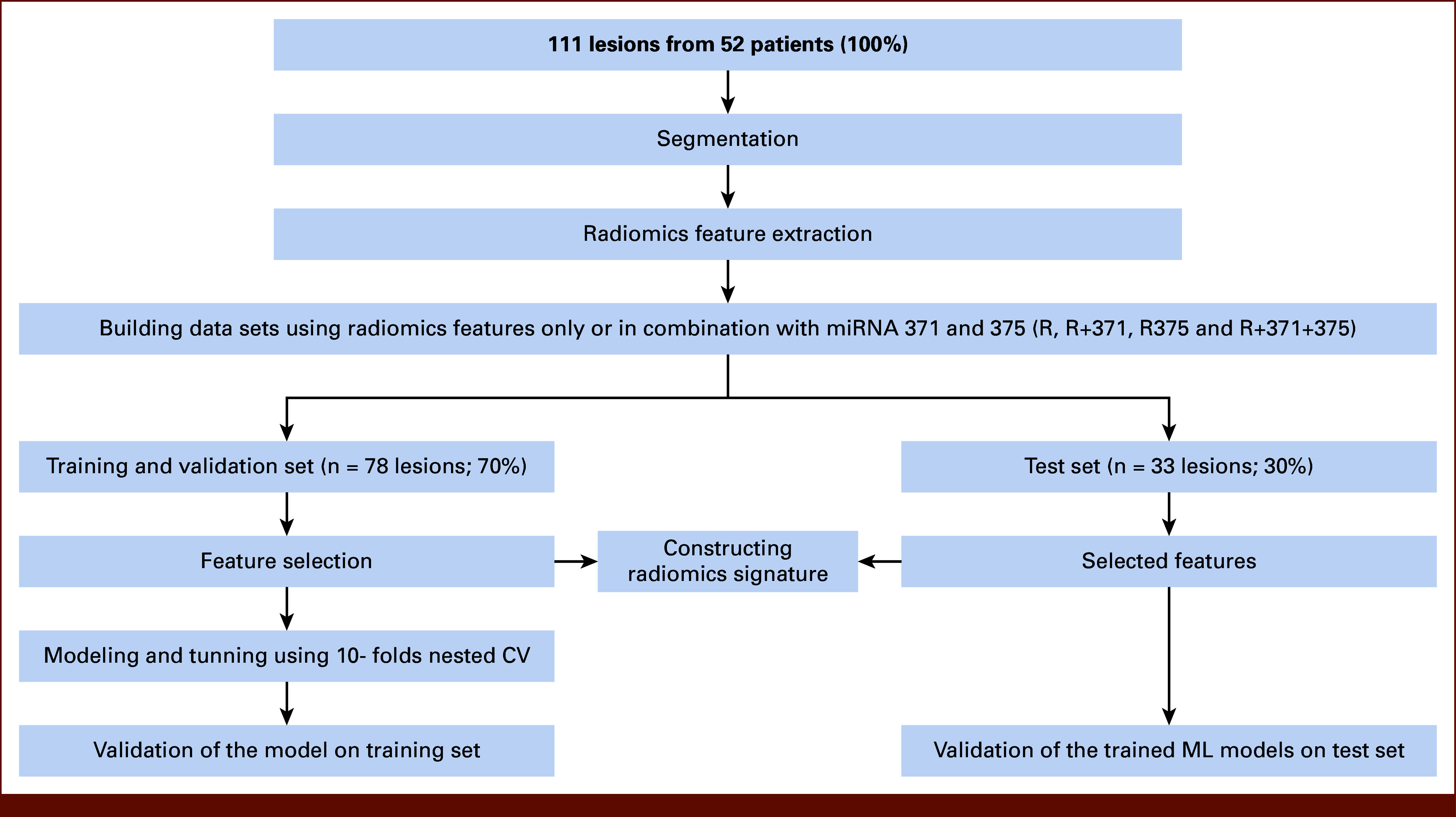
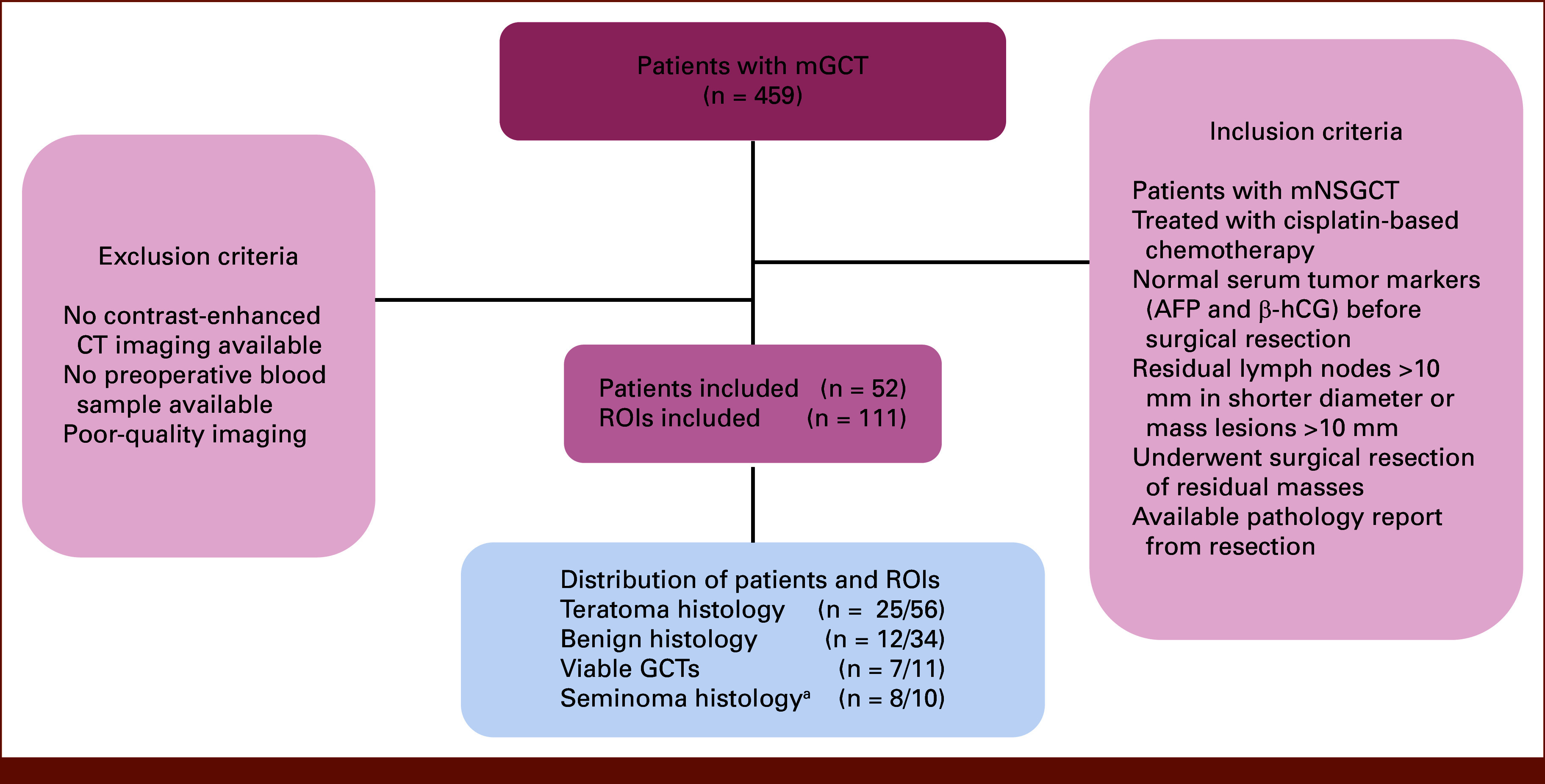
Methods: We retrospectively identified 111 lesions, divided into training and test sets (n = 78 v 33) with equal class distribution. 3D Slicer was used to segment lesions with a short axis of >10 mm from the postchemo-presurgical CT images, and radiomics features were extracted. Presurgery plasma miR371-375 levels were measured by real-time polymerase chain reaction. Four machine learning models evaluated the predictive value of radiomics alone (R-only) and combined with miR371-375 levels, and the best performer was selected. Clinical factors associated with teratoma from univariate analysis were included in multivariate analysis with the best radiomics signature to assess their impact on predicting teratoma histology.
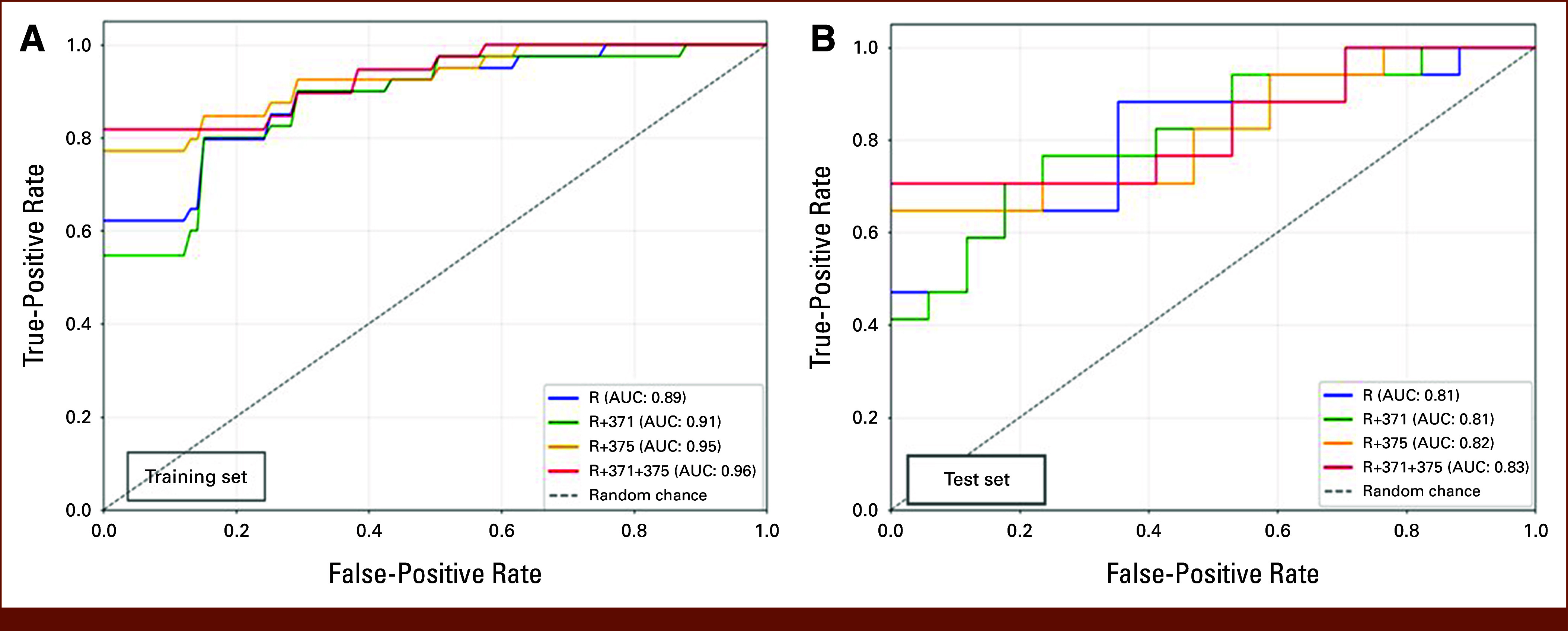
Results: The CatBoost (CB) model R + 371 + 375 exhibited the best and most robust overall accuracy for predicting residual teratoma, with the highest AUC values (0.96, 95% CI, 0.88 to 1.0 for training, 0.83, 95% CI, 0.68 to 0.98 for testing) and a well-balanced sensitivity and specificity. Univariate analysis identified presurgery alpha-fetoprotein (P = .01), beta-human chorionic gonadotropin (P = .01), initial teratoma pathology (P = .01), and lymph node metastases (P = .02) as significant predictors for teratoma. Multivariate analysis included these features and the radiomics signature, which was the strongest independent predictor (P < .0001).
Conclusion: Combining miR371-375 with CT radiomics features improves the accuracy of predicting teratoma histology of postchemotherapy residual disease in mNSGCTs and, therefore, has the potential to guide treatment decision making.

 求助内容:
求助内容: 应助结果提醒方式:


