Yeon-Koo Kang, Jae Won Min, Soo Jin Kwon, Seunggyun Ha
{"title":"Reliability of Automated Amyloid PET Quantification: Real-World Validation of Commercial Tools Against Centiloid Project Method.","authors":"Yeon-Koo Kang, Jae Won Min, Soo Jin Kwon, Seunggyun Ha","doi":"10.3390/tomography11080086","DOIUrl":null,"url":null,"abstract":"<p><p><b>Background:</b> Despite the growing demand for amyloid PET quantification, practical challenges remain. As automated software platforms are increasingly adopted to address these limitations, we evaluated the reliability of commercial tools for Centiloid quantification against the original Centiloid Project method. <b>Methods:</b> This retrospective study included 332 amyloid PET scans (165 [<sup>18</sup>F]Florbetaben; 167 [<sup>18</sup>F]Flutemetamol) performed for suspected mild cognitive impairments or dementia, paired with T1-weighted MRI within one year. Centiloid values were calculated using three automated software platforms, BTXBrain, MIMneuro, and SCALE PET, and compared with the original Centiloid method. The agreement was assessed using Pearson's correlation coefficient, the intraclass correlation coefficient (ICC), a Passing-Bablok regression, and Bland-Altman plots. The concordance with the visual interpretation was evaluated using receiver operating characteristic (ROC) curves. <b>Results:</b> BTXBrain (R = 0.993; ICC = 0.986) and SCALE PET (R = 0.992; ICC = 0.991) demonstrated an excellent correlation with the reference, while MIMneuro showed a slightly lower agreement (R = 0.974; ICC = 0.966). BTXBrain exhibited a proportional underestimation (slope = 0.872 [0.860-0.885]), MIMneuro showed a significant overestimation (slope = 1.053 [1.026-1.081]), and SCALE PET demonstrated a minimal bias (slope = 1.014 [0.999-1.029]). The bias pattern was particularly noted for FMM. All platforms maintained their trends for correlations and biases when focusing on subthreshold-to-low-positive ranges (0-50 Centiloid units). However, all platforms showed an excellent agreement with the visual interpretation (areas under ROC curves > 0.996 for all). <b>Conclusions:</b> Three automated platforms demonstrated an acceptable reliability for Centiloid quantification, although software-specific biases were observed. These differences did not impair their feasibility in aiding the image interpretation, as supported by the concordance with visual readings. Nevertheless, users should recognize the platform-specific characteristics when applying diagnostic thresholds or interpreting longitudinal changes.</p>","PeriodicalId":51330,"journal":{"name":"Tomography","volume":"11 8","pages":""},"PeriodicalIF":2.2000,"publicationDate":"2025-07-30","publicationTypes":"Journal Article","fieldsOfStudy":null,"isOpenAccess":false,"openAccessPdf":"https://www.ncbi.nlm.nih.gov/pmc/articles/PMC12389775/pdf/","citationCount":"0","resultStr":null,"platform":"Semanticscholar","paperid":null,"PeriodicalName":"Tomography","FirstCategoryId":"3","ListUrlMain":"https://doi.org/10.3390/tomography11080086","RegionNum":4,"RegionCategory":"医学","ArticlePicture":[],"TitleCN":null,"AbstractTextCN":null,"PMCID":null,"EPubDate":"","PubModel":"","JCR":"Q2","JCRName":"RADIOLOGY, NUCLEAR MEDICINE & MEDICAL IMAGING","Score":null,"Total":0}
引用次数: 0
Abstract
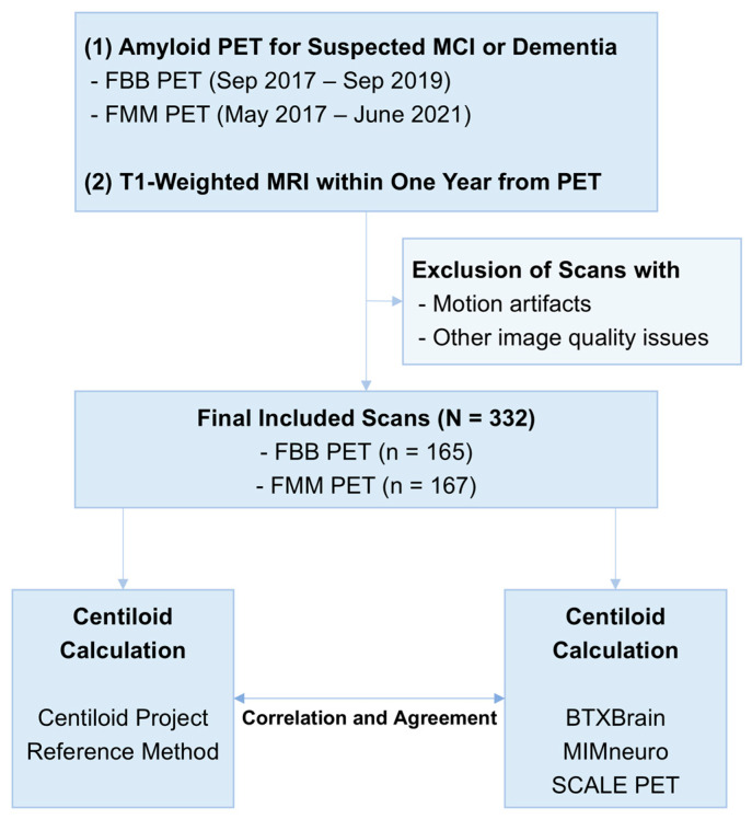
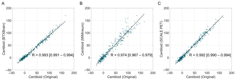
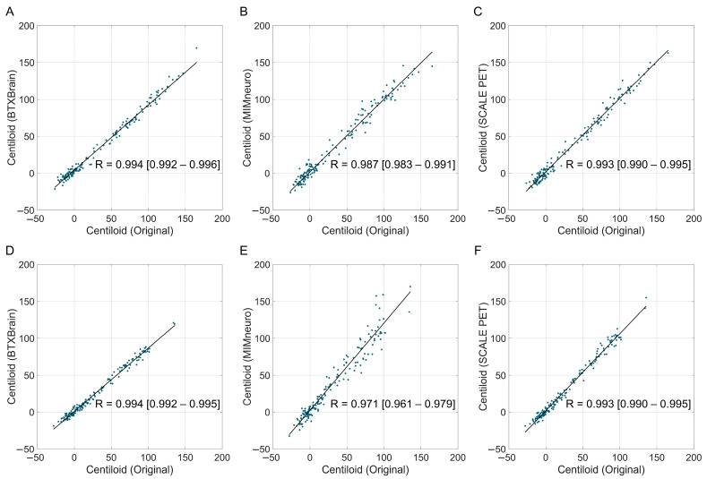
Background: Despite the growing demand for amyloid PET quantification, practical challenges remain. As automated software platforms are increasingly adopted to address these limitations, we evaluated the reliability of commercial tools for Centiloid quantification against the original Centiloid Project method. Methods: This retrospective study included 332 amyloid PET scans (165 [18F]Florbetaben; 167 [18F]Flutemetamol) performed for suspected mild cognitive impairments or dementia, paired with T1-weighted MRI within one year. Centiloid values were calculated using three automated software platforms, BTXBrain, MIMneuro, and SCALE PET, and compared with the original Centiloid method. The agreement was assessed using Pearson's correlation coefficient, the intraclass correlation coefficient (ICC), a Passing-Bablok regression, and Bland-Altman plots. The concordance with the visual interpretation was evaluated using receiver operating characteristic (ROC) curves. Results: BTXBrain (R = 0.993; ICC = 0.986) and SCALE PET (R = 0.992; ICC = 0.991) demonstrated an excellent correlation with the reference, while MIMneuro showed a slightly lower agreement (R = 0.974; ICC = 0.966). BTXBrain exhibited a proportional underestimation (slope = 0.872 [0.860-0.885]), MIMneuro showed a significant overestimation (slope = 1.053 [1.026-1.081]), and SCALE PET demonstrated a minimal bias (slope = 1.014 [0.999-1.029]). The bias pattern was particularly noted for FMM. All platforms maintained their trends for correlations and biases when focusing on subthreshold-to-low-positive ranges (0-50 Centiloid units). However, all platforms showed an excellent agreement with the visual interpretation (areas under ROC curves > 0.996 for all). Conclusions: Three automated platforms demonstrated an acceptable reliability for Centiloid quantification, although software-specific biases were observed. These differences did not impair their feasibility in aiding the image interpretation, as supported by the concordance with visual readings. Nevertheless, users should recognize the platform-specific characteristics when applying diagnostic thresholds or interpreting longitudinal changes.
TomographyMedicine-Radiology, Nuclear Medicine and Imaging
CiteScore
2.70
自引率
10.50%
发文量
222
期刊介绍:
TomographyTM publishes basic (technical and pre-clinical) and clinical scientific articles which involve the advancement of imaging technologies. Tomography encompasses studies that use single or multiple imaging modalities including for example CT, US, PET, SPECT, MR and hyperpolarization technologies, as well as optical modalities (i.e. bioluminescence, photoacoustic, endomicroscopy, fiber optic imaging and optical computed tomography) in basic sciences, engineering, preclinical and clinical medicine.
Tomography also welcomes studies involving exploration and refinement of contrast mechanisms and image-derived metrics within and across modalities toward the development of novel imaging probes for image-based feedback and intervention. The use of imaging in biology and medicine provides unparalleled opportunities to noninvasively interrogate tissues to obtain real-time dynamic and quantitative information required for diagnosis and response to interventions and to follow evolving pathological conditions. As multi-modal studies and the complexities of imaging technologies themselves are ever increasing to provide advanced information to scientists and clinicians.
Tomography provides a unique publication venue allowing investigators the opportunity to more precisely communicate integrated findings related to the diverse and heterogeneous features associated with underlying anatomical, physiological, functional, metabolic and molecular genetic activities of normal and diseased tissue. Thus Tomography publishes peer-reviewed articles which involve the broad use of imaging of any tissue and disease type including both preclinical and clinical investigations. In addition, hardware/software along with chemical and molecular probe advances are welcome as they are deemed to significantly contribute towards the long-term goal of improving the overall impact of imaging on scientific and clinical discovery.

 求助内容:
求助内容: 应助结果提醒方式:


