Roseline Oluwaseun Ogundokun, Pius Adewale Owolawi, Chunling Tu, Etienne van Wyk
{"title":"Autoencoder-Assisted Stacked Ensemble Learning for Lymphoma Subtype Classification: A Hybrid Deep Learning and Machine Learning Approach.","authors":"Roseline Oluwaseun Ogundokun, Pius Adewale Owolawi, Chunling Tu, Etienne van Wyk","doi":"10.3390/tomography11080091","DOIUrl":null,"url":null,"abstract":"<p><strong>Background: </strong>Accurate subtype identification of lymphoma cancer is crucial for effective diagnosis and treatment planning. Although standard deep learning algorithms have demonstrated robustness, they are still prone to overfitting and limited generalization, necessitating more reliable and robust methods.</p><p><strong>Objectives: </strong>This study presents an autoencoder-augmented stacked ensemble learning (SEL) framework integrating deep feature extraction (DFE) and ensembles of machine learning classifiers to improve lymphoma subtype identification.</p><p><strong>Methods: </strong>Convolutional autoencoder (CAE) was utilized to obtain high-level feature representations of histopathological images, followed by dimensionality reduction via Principal Component Analysis (PCA). Various models were utilized for classifying extracted features, i.e., Random Forest (RF), Support Vector Machine (SVM), Multi-Layer Perceptron (MLP), AdaBoost, and Extra Trees classifiers. A Gradient Boosting Machine (GBM) meta-classifier was utilized in an SEL approach to further fine-tune final predictions.</p><p><strong>Results: </strong>All the models were tested using accuracy, area under the curve (AUC), and Average Precision (AP) metrics. The stacked ensemble classifier performed better than all the individual models with a 99.04% accuracy, 0.9998 AUC, and 0.9996 AP, far exceeding what regular deep learning (DL) methods would achieve. Of standalone classifiers, MLP (97.71% accuracy, 0.9986 AUC, 0.9973 AP) and Random Forest (96.71% accuracy, 0.9977 AUC, 0.9953 AP) provided the best prediction performance, while AdaBoost was the poorest performer (68.25% accuracy, 0.8194 AUC, 0.6424 AP). PCA and t-SNE plots confirmed that DFE effectively enhances class discrimination.</p><p><strong>Conclusion: </strong>This study demonstrates a highly accurate and reliable approach to lymphoma classification by using autoencoder-assisted ensemble learning, reducing the misclassification rate and significantly enhancing the accuracy of diagnosis. AI-based models are designed to assist pathologists by providing interpretable outputs such as class probabilities and visualizations (e.g., Grad-CAM), enabling them to understand and validate predictions in the diagnostic workflow. Future studies should enhance computational efficacy and conduct multi-centre validation studies to confirm the model's generalizability on extensive collections of histopathological datasets.</p>","PeriodicalId":51330,"journal":{"name":"Tomography","volume":"11 8","pages":""},"PeriodicalIF":2.2000,"publicationDate":"2025-08-18","publicationTypes":"Journal Article","fieldsOfStudy":null,"isOpenAccess":false,"openAccessPdf":"https://www.ncbi.nlm.nih.gov/pmc/articles/PMC12389832/pdf/","citationCount":"0","resultStr":null,"platform":"Semanticscholar","paperid":null,"PeriodicalName":"Tomography","FirstCategoryId":"3","ListUrlMain":"https://doi.org/10.3390/tomography11080091","RegionNum":4,"RegionCategory":"医学","ArticlePicture":[],"TitleCN":null,"AbstractTextCN":null,"PMCID":null,"EPubDate":"","PubModel":"","JCR":"Q2","JCRName":"RADIOLOGY, NUCLEAR MEDICINE & MEDICAL IMAGING","Score":null,"Total":0}
引用次数: 0
Abstract
Background: Accurate subtype identification of lymphoma cancer is crucial for effective diagnosis and treatment planning. Although standard deep learning algorithms have demonstrated robustness, they are still prone to overfitting and limited generalization, necessitating more reliable and robust methods.
Objectives: This study presents an autoencoder-augmented stacked ensemble learning (SEL) framework integrating deep feature extraction (DFE) and ensembles of machine learning classifiers to improve lymphoma subtype identification.
Methods: Convolutional autoencoder (CAE) was utilized to obtain high-level feature representations of histopathological images, followed by dimensionality reduction via Principal Component Analysis (PCA). Various models were utilized for classifying extracted features, i.e., Random Forest (RF), Support Vector Machine (SVM), Multi-Layer Perceptron (MLP), AdaBoost, and Extra Trees classifiers. A Gradient Boosting Machine (GBM) meta-classifier was utilized in an SEL approach to further fine-tune final predictions.
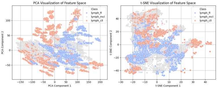
Results: All the models were tested using accuracy, area under the curve (AUC), and Average Precision (AP) metrics. The stacked ensemble classifier performed better than all the individual models with a 99.04% accuracy, 0.9998 AUC, and 0.9996 AP, far exceeding what regular deep learning (DL) methods would achieve. Of standalone classifiers, MLP (97.71% accuracy, 0.9986 AUC, 0.9973 AP) and Random Forest (96.71% accuracy, 0.9977 AUC, 0.9953 AP) provided the best prediction performance, while AdaBoost was the poorest performer (68.25% accuracy, 0.8194 AUC, 0.6424 AP). PCA and t-SNE plots confirmed that DFE effectively enhances class discrimination.
Conclusion: This study demonstrates a highly accurate and reliable approach to lymphoma classification by using autoencoder-assisted ensemble learning, reducing the misclassification rate and significantly enhancing the accuracy of diagnosis. AI-based models are designed to assist pathologists by providing interpretable outputs such as class probabilities and visualizations (e.g., Grad-CAM), enabling them to understand and validate predictions in the diagnostic workflow. Future studies should enhance computational efficacy and conduct multi-centre validation studies to confirm the model's generalizability on extensive collections of histopathological datasets.
TomographyMedicine-Radiology, Nuclear Medicine and Imaging
CiteScore
2.70
自引率
10.50%
发文量
222
期刊介绍:
TomographyTM publishes basic (technical and pre-clinical) and clinical scientific articles which involve the advancement of imaging technologies. Tomography encompasses studies that use single or multiple imaging modalities including for example CT, US, PET, SPECT, MR and hyperpolarization technologies, as well as optical modalities (i.e. bioluminescence, photoacoustic, endomicroscopy, fiber optic imaging and optical computed tomography) in basic sciences, engineering, preclinical and clinical medicine.
Tomography also welcomes studies involving exploration and refinement of contrast mechanisms and image-derived metrics within and across modalities toward the development of novel imaging probes for image-based feedback and intervention. The use of imaging in biology and medicine provides unparalleled opportunities to noninvasively interrogate tissues to obtain real-time dynamic and quantitative information required for diagnosis and response to interventions and to follow evolving pathological conditions. As multi-modal studies and the complexities of imaging technologies themselves are ever increasing to provide advanced information to scientists and clinicians.
Tomography provides a unique publication venue allowing investigators the opportunity to more precisely communicate integrated findings related to the diverse and heterogeneous features associated with underlying anatomical, physiological, functional, metabolic and molecular genetic activities of normal and diseased tissue. Thus Tomography publishes peer-reviewed articles which involve the broad use of imaging of any tissue and disease type including both preclinical and clinical investigations. In addition, hardware/software along with chemical and molecular probe advances are welcome as they are deemed to significantly contribute towards the long-term goal of improving the overall impact of imaging on scientific and clinical discovery.

 求助内容:
求助内容: 应助结果提醒方式:


