Lauren Brenner, Tanner H Robison, Timothy D Johnson, Kristen Pettit, Moshe Talpaz, Thomas L Chenevert, Brian D Ross, Gary D Luker
{"title":"Fat Fraction MRI for Longitudinal Assessment of Bone Marrow Heterogeneity in a Mouse Model of Myelofibrosis.","authors":"Lauren Brenner, Tanner H Robison, Timothy D Johnson, Kristen Pettit, Moshe Talpaz, Thomas L Chenevert, Brian D Ross, Gary D Luker","doi":"10.3390/tomography11080082","DOIUrl":null,"url":null,"abstract":"<p><strong>Background/objectives: </strong>Myelofibrosis (MF) is a myeloproliferative neoplasm characterized by the replacement of healthy bone marrow (BM) with malignant and fibrotic tissue. In a healthy state, bone marrow is composed of approximately 60-70% fat cells, which are replaced as disease progresses. Proton density fat fraction (PDFF), a non-invasive and quantitative MRI metric, enables analysis of BM architecture by measuring the percentage of fat versus cells in the environment. Our objective is to investigate variance in quantitative PDFF-MRI values over time as a marker of disease progression and response to treatment.</p><p><strong>Methods: </strong>We analyzed existing data from three cohorts of mice: two groups with MF that failed to respond to therapy with approved drugs for MF (ruxolitinib, fedratinib), investigational compounds (navitoclax, balixafortide), or vehicle and monitored over time by MRI; the third group consisted of healthy controls imaged at a single time point. Using in-house MATLAB programs, we performed a voxel-wise analysis of PDFF values in lower extremity bone marrow, specifically comparing the variance of each voxel within and among mice.</p><p><strong>Results: </strong>Our findings revealed a significant difference in PDFF values between healthy and diseased BM. With progressive disease non-responsive to therapy, the expansion of hematopoietic cells in BM nearly completely replaced normal fat, as determined by a markedly reduced PDFF and notable reduction in the variance in PDFF values in bone marrow over time.</p><p><strong>Conclusions: </strong>This study validated our hypothesis that the variance in PDFF in BM decreases with disease progression, indicating pathologic expansion of hematopoietic cells. We can conclude that disease progression can be tracked by a decrease in PDFF values. Analyzing variance in PDFF may improve the assessment of disease progression in pre-clinical models and ultimately patients with MF.</p>","PeriodicalId":51330,"journal":{"name":"Tomography","volume":"11 8","pages":""},"PeriodicalIF":2.2000,"publicationDate":"2025-07-28","publicationTypes":"Journal Article","fieldsOfStudy":null,"isOpenAccess":false,"openAccessPdf":"https://www.ncbi.nlm.nih.gov/pmc/articles/PMC12390422/pdf/","citationCount":"0","resultStr":null,"platform":"Semanticscholar","paperid":null,"PeriodicalName":"Tomography","FirstCategoryId":"3","ListUrlMain":"https://doi.org/10.3390/tomography11080082","RegionNum":4,"RegionCategory":"医学","ArticlePicture":[],"TitleCN":null,"AbstractTextCN":null,"PMCID":null,"EPubDate":"","PubModel":"","JCR":"Q2","JCRName":"RADIOLOGY, NUCLEAR MEDICINE & MEDICAL IMAGING","Score":null,"Total":0}
引用次数: 0
Abstract
Background/objectives: Myelofibrosis (MF) is a myeloproliferative neoplasm characterized by the replacement of healthy bone marrow (BM) with malignant and fibrotic tissue. In a healthy state, bone marrow is composed of approximately 60-70% fat cells, which are replaced as disease progresses. Proton density fat fraction (PDFF), a non-invasive and quantitative MRI metric, enables analysis of BM architecture by measuring the percentage of fat versus cells in the environment. Our objective is to investigate variance in quantitative PDFF-MRI values over time as a marker of disease progression and response to treatment.
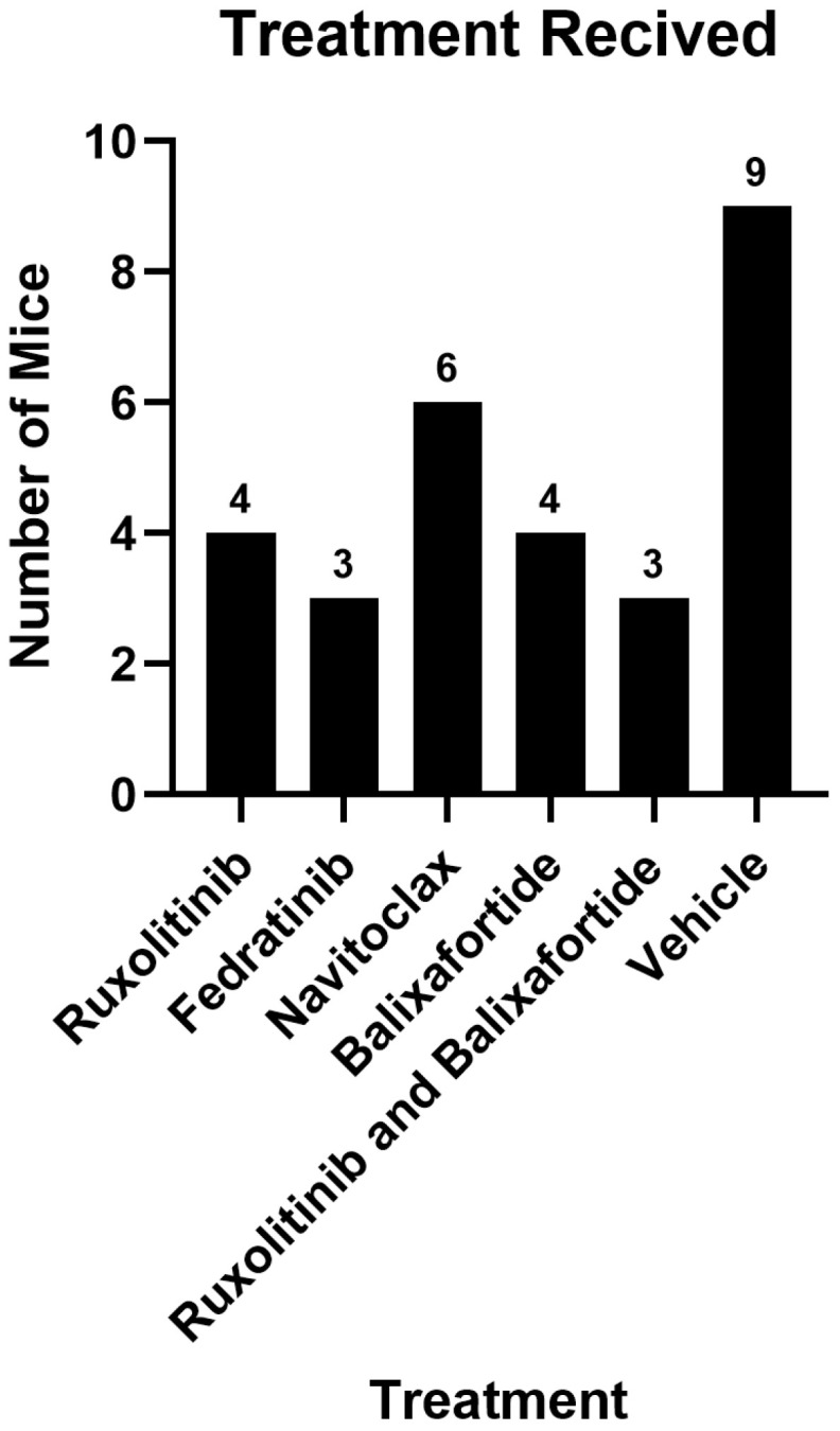
Methods: We analyzed existing data from three cohorts of mice: two groups with MF that failed to respond to therapy with approved drugs for MF (ruxolitinib, fedratinib), investigational compounds (navitoclax, balixafortide), or vehicle and monitored over time by MRI; the third group consisted of healthy controls imaged at a single time point. Using in-house MATLAB programs, we performed a voxel-wise analysis of PDFF values in lower extremity bone marrow, specifically comparing the variance of each voxel within and among mice.
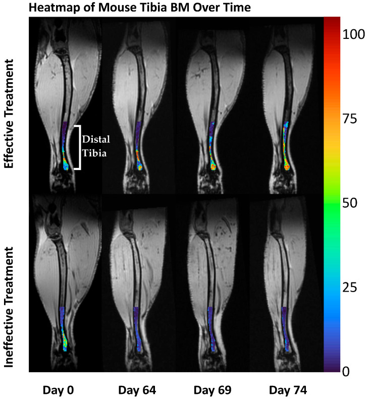
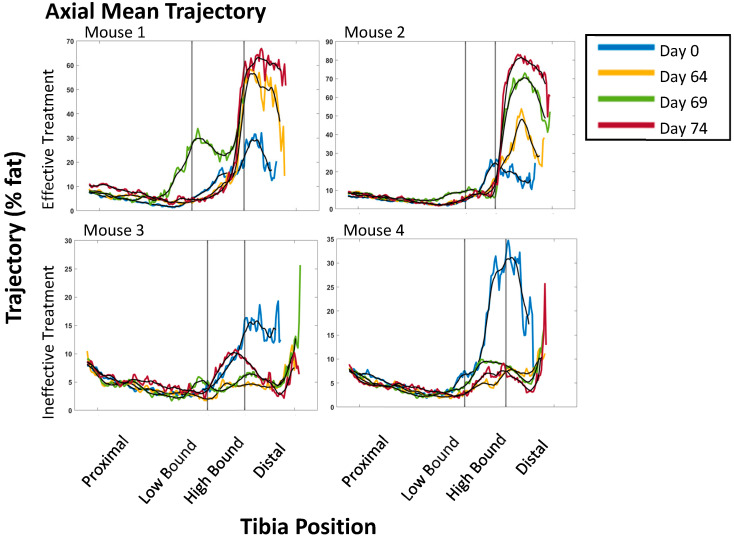
Results: Our findings revealed a significant difference in PDFF values between healthy and diseased BM. With progressive disease non-responsive to therapy, the expansion of hematopoietic cells in BM nearly completely replaced normal fat, as determined by a markedly reduced PDFF and notable reduction in the variance in PDFF values in bone marrow over time.
Conclusions: This study validated our hypothesis that the variance in PDFF in BM decreases with disease progression, indicating pathologic expansion of hematopoietic cells. We can conclude that disease progression can be tracked by a decrease in PDFF values. Analyzing variance in PDFF may improve the assessment of disease progression in pre-clinical models and ultimately patients with MF.
TomographyMedicine-Radiology, Nuclear Medicine and Imaging
CiteScore
2.70
自引率
10.50%
发文量
222
期刊介绍:
TomographyTM publishes basic (technical and pre-clinical) and clinical scientific articles which involve the advancement of imaging technologies. Tomography encompasses studies that use single or multiple imaging modalities including for example CT, US, PET, SPECT, MR and hyperpolarization technologies, as well as optical modalities (i.e. bioluminescence, photoacoustic, endomicroscopy, fiber optic imaging and optical computed tomography) in basic sciences, engineering, preclinical and clinical medicine.
Tomography also welcomes studies involving exploration and refinement of contrast mechanisms and image-derived metrics within and across modalities toward the development of novel imaging probes for image-based feedback and intervention. The use of imaging in biology and medicine provides unparalleled opportunities to noninvasively interrogate tissues to obtain real-time dynamic and quantitative information required for diagnosis and response to interventions and to follow evolving pathological conditions. As multi-modal studies and the complexities of imaging technologies themselves are ever increasing to provide advanced information to scientists and clinicians.
Tomography provides a unique publication venue allowing investigators the opportunity to more precisely communicate integrated findings related to the diverse and heterogeneous features associated with underlying anatomical, physiological, functional, metabolic and molecular genetic activities of normal and diseased tissue. Thus Tomography publishes peer-reviewed articles which involve the broad use of imaging of any tissue and disease type including both preclinical and clinical investigations. In addition, hardware/software along with chemical and molecular probe advances are welcome as they are deemed to significantly contribute towards the long-term goal of improving the overall impact of imaging on scientific and clinical discovery.

 求助内容:
求助内容: 应助结果提醒方式:


