{"title":"Deep Learning-Based Nuclei Segmentation and Melanoma Detection in Skin Histopathological Image Using Test Image Augmentation and Ensemble Model.","authors":"Mohammadesmaeil Akbarpour, Hamed Fazlollahiaghamalek, Mahdi Barati, Mehrdad Hashemi Kamangar, Mrinal Mandal","doi":"10.3390/jimaging11080274","DOIUrl":null,"url":null,"abstract":"<p><p>Histopathological images play a crucial role in diagnosing skin cancer. However, due to the very large size of digital histopathological images (typically in the order of billion pixels), manual image analysis is tedious and time-consuming. Therefore, there has been significant interest in developing Artificial Intelligence (AI)-enabled computer-aided diagnosis (CAD) techniques for skin cancer detection. Due to the diversity of uncertain cell boundaries, automated nuclei segmentation of histopathological images remains challenging. Automating the identification of abnormal cell nuclei and analyzing their distribution across multiple tissue sections can significantly expedite comprehensive diagnostic assessments. In this paper, a deep neural network (DNN)-based technique is proposed to segment nuclei and detect melanoma in histopathological images. To achieve a robust performance, a test image is first augmented by various geometric operations. The augmented images are then passed through the DNN and the individual outputs are combined to obtain the final nuclei-segmented image. A morphological technique is then applied on the nuclei-segmented image to detect the melanoma region in the image. Experimental results show that the proposed technique can achieve a Dice score of 91.61% and 87.9% for nuclei segmentation and melanoma detection, respectively.</p>","PeriodicalId":37035,"journal":{"name":"Journal of Imaging","volume":"11 8","pages":""},"PeriodicalIF":2.7000,"publicationDate":"2025-08-15","publicationTypes":"Journal Article","fieldsOfStudy":null,"isOpenAccess":false,"openAccessPdf":"https://www.ncbi.nlm.nih.gov/pmc/articles/PMC12387607/pdf/","citationCount":"0","resultStr":null,"platform":"Semanticscholar","paperid":null,"PeriodicalName":"Journal of Imaging","FirstCategoryId":"1085","ListUrlMain":"https://doi.org/10.3390/jimaging11080274","RegionNum":0,"RegionCategory":null,"ArticlePicture":[],"TitleCN":null,"AbstractTextCN":null,"PMCID":null,"EPubDate":"","PubModel":"","JCR":"Q3","JCRName":"IMAGING SCIENCE & PHOTOGRAPHIC TECHNOLOGY","Score":null,"Total":0}
引用次数: 0
Abstract
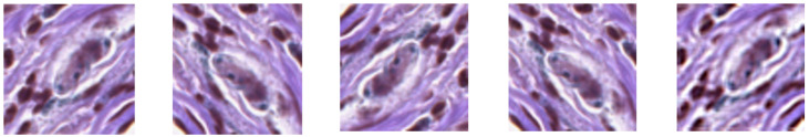
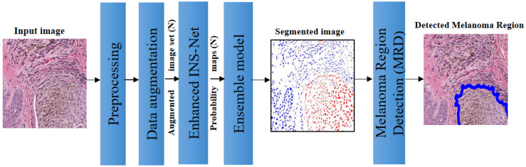
Histopathological images play a crucial role in diagnosing skin cancer. However, due to the very large size of digital histopathological images (typically in the order of billion pixels), manual image analysis is tedious and time-consuming. Therefore, there has been significant interest in developing Artificial Intelligence (AI)-enabled computer-aided diagnosis (CAD) techniques for skin cancer detection. Due to the diversity of uncertain cell boundaries, automated nuclei segmentation of histopathological images remains challenging. Automating the identification of abnormal cell nuclei and analyzing their distribution across multiple tissue sections can significantly expedite comprehensive diagnostic assessments. In this paper, a deep neural network (DNN)-based technique is proposed to segment nuclei and detect melanoma in histopathological images. To achieve a robust performance, a test image is first augmented by various geometric operations. The augmented images are then passed through the DNN and the individual outputs are combined to obtain the final nuclei-segmented image. A morphological technique is then applied on the nuclei-segmented image to detect the melanoma region in the image. Experimental results show that the proposed technique can achieve a Dice score of 91.61% and 87.9% for nuclei segmentation and melanoma detection, respectively.

 求助内容:
求助内容: 应助结果提醒方式:


