Gauri Darekar, Taslim Murad, Hui-Yuan Miao, Deepa S Thakuri, Ganesh B Chand
{"title":"An explainable AI approach for mapping multivariate regional brain age and clinical severity patterns in Alzheimer's disease.","authors":"Gauri Darekar, Taslim Murad, Hui-Yuan Miao, Deepa S Thakuri, Ganesh B Chand","doi":"10.1093/biomethods/bpaf051","DOIUrl":null,"url":null,"abstract":"<p><p>Age is a significant risk factor for mild cognitive impairment (MCI) and Alzheimer's disease (AD) and identifying brain age patterns is critical for comprehending the normal aging and MCI/AD processes. Prior studies have widely established the univariate relationships between brain regions and age, while multivariate associations remain largely unexplored. Herein, various artificial intelligence (AI) models were used to perform brain age prediction using an MRI dataset (<i>n</i> = 825). The optimal AI model was then integrated with the feature importance methods, namely Shapley additive explanations (SHAP), local interpretable model-agnostic explanations, and layer-wise relevance propagation, to identify the significant multivariate brain regions hierarchically involved in this prediction. Our results showed that the deep learning model (referred to as AgeNet) outperformed conventional machine learning models for brain age prediction, and that AgeNet integrated with SHAP (referred to as AgeNet-SHAP) identified all ground-truth perturbed regions as key predictors of brain age in semi-simulation, demonstrating the validity of our methodology. In the experimental dataset, when compared to cognitively normal (CN) participants, MCI exhibited moderate differences in brain regions, whereas AD showed highly robust and widely distributed regional differences. Individualized AgeNet-SHAP regional features further showed associations with clinical severity scores in the AD continuum. These results collectively facilitate data-driven explainable AI approaches for disease progression, diagnostics, prognostics, and personalized medicine efforts.</p>","PeriodicalId":36528,"journal":{"name":"Biology Methods and Protocols","volume":"10 1","pages":"bpaf051"},"PeriodicalIF":1.3000,"publicationDate":"2025-08-07","publicationTypes":"Journal Article","fieldsOfStudy":null,"isOpenAccess":false,"openAccessPdf":"https://www.ncbi.nlm.nih.gov/pmc/articles/PMC12377905/pdf/","citationCount":"0","resultStr":null,"platform":"Semanticscholar","paperid":null,"PeriodicalName":"Biology Methods and Protocols","FirstCategoryId":"1085","ListUrlMain":"https://doi.org/10.1093/biomethods/bpaf051","RegionNum":0,"RegionCategory":null,"ArticlePicture":[],"TitleCN":null,"AbstractTextCN":null,"PMCID":null,"EPubDate":"2025/1/1 0:00:00","PubModel":"eCollection","JCR":"Q3","JCRName":"BIOCHEMICAL RESEARCH METHODS","Score":null,"Total":0}
引用次数: 0
Abstract
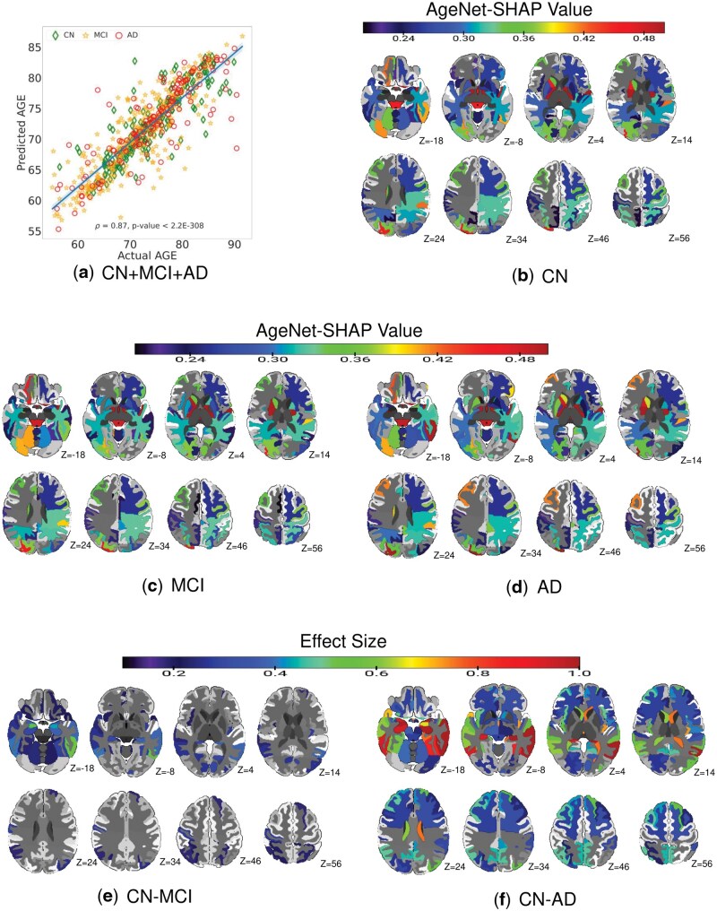
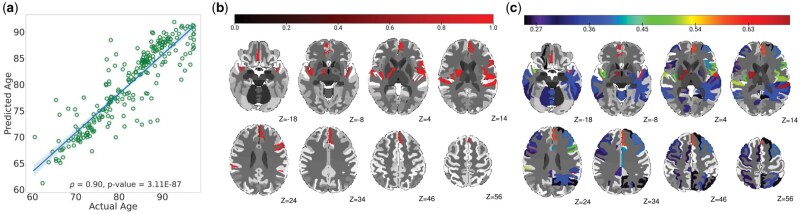
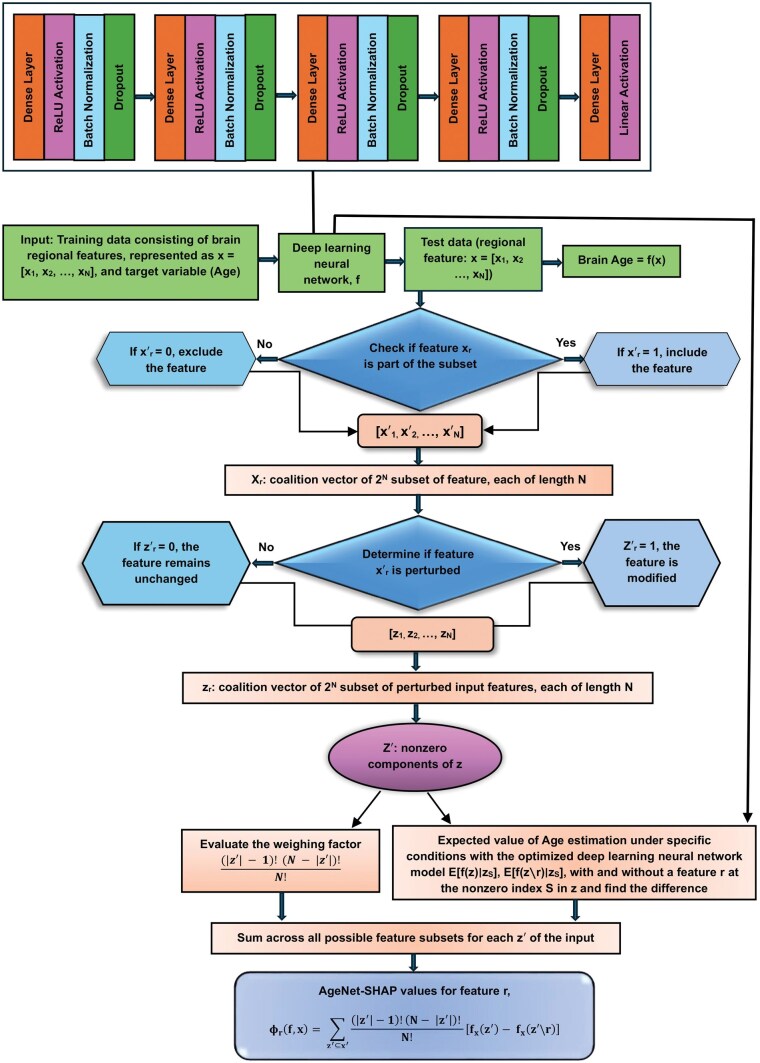
Age is a significant risk factor for mild cognitive impairment (MCI) and Alzheimer's disease (AD) and identifying brain age patterns is critical for comprehending the normal aging and MCI/AD processes. Prior studies have widely established the univariate relationships between brain regions and age, while multivariate associations remain largely unexplored. Herein, various artificial intelligence (AI) models were used to perform brain age prediction using an MRI dataset (n = 825). The optimal AI model was then integrated with the feature importance methods, namely Shapley additive explanations (SHAP), local interpretable model-agnostic explanations, and layer-wise relevance propagation, to identify the significant multivariate brain regions hierarchically involved in this prediction. Our results showed that the deep learning model (referred to as AgeNet) outperformed conventional machine learning models for brain age prediction, and that AgeNet integrated with SHAP (referred to as AgeNet-SHAP) identified all ground-truth perturbed regions as key predictors of brain age in semi-simulation, demonstrating the validity of our methodology. In the experimental dataset, when compared to cognitively normal (CN) participants, MCI exhibited moderate differences in brain regions, whereas AD showed highly robust and widely distributed regional differences. Individualized AgeNet-SHAP regional features further showed associations with clinical severity scores in the AD continuum. These results collectively facilitate data-driven explainable AI approaches for disease progression, diagnostics, prognostics, and personalized medicine efforts.

 求助内容:
求助内容: 应助结果提醒方式:


