S Q Qu, N N Liu, T J Qin, Z F Xu, B Li, L J Pan, M Jiao, Q Y Gao, H J Wang, X F Ai, Z J Xiao
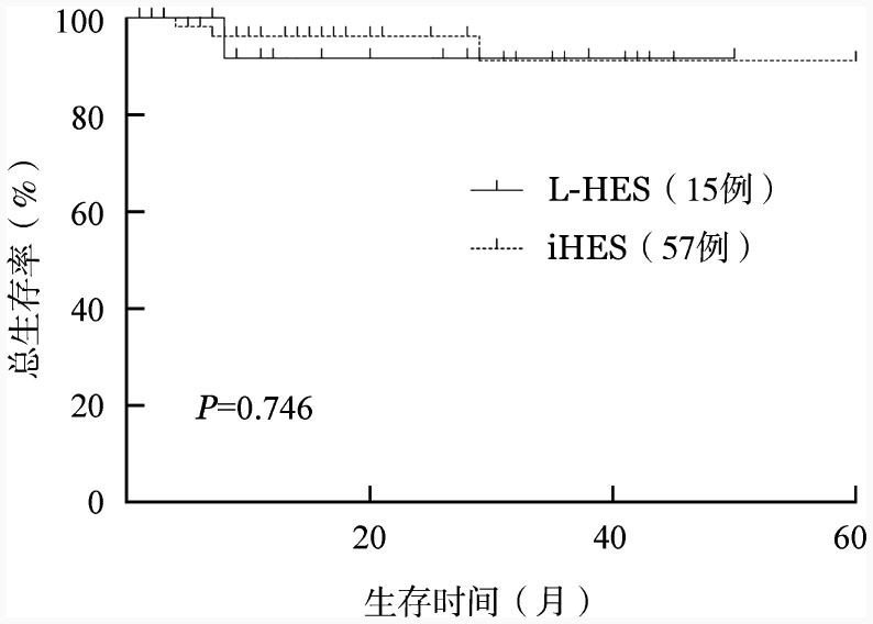
{"title":"[Efficacy and survival outcomes of patients with lymphocytic variant hypereosinophilic syndrome].","authors":"S Q Qu, N N Liu, T J Qin, Z F Xu, B Li, L J Pan, M Jiao, Q Y Gao, H J Wang, X F Ai, Z J Xiao","doi":"10.3760/cma.j.cn121090-20250109-00019","DOIUrl":null,"url":null,"abstract":"<p><p><b>Objective:</b> To analyze the clinical characteristics, therapeutic responses, and survival outcomes of patients with lymphocytic variant hypereosinophilic syndrome (L-HES) . <b>Methods:</b> We retrospectively reviewed clinical data from 16 consecutive patients diagnosed with L-HES at the Institute of Hematology and Blood Diseases Hospital, Chinese Academy of Medical Sciences, between July 2019 and October 2024. A control group of 65 patients with idiopathic hypereosinophilic syndrome (iHES), diagnosed during the same period, was used for comparison. Clinical and laboratory characteristics, therapeutic responses, and survival outcomes were compared between the two groups. <b>Results:</b> The most frequently involved organs at presentation in patients with L-HES were the skin (75.0%), gastrointestinal tract (25.0%), respiratory tract (18.8%), lymph nodes (18.8%), heart (12.5%), and spleen (6.3%). Compared with iHES patients, patients with L-HES had a significantly higher incidence of skin involvement (<i>P</i>=0.016), with no statistically significant differences observed in the involvement of other organs. No statistically significant differences were found in complete blood count parameters between the two groups. Multiparameter flow cytometry revealed that the median percentage of CD3(-)CD4(+) T cells in the peripheral blood of patients with L-HES was 4.08% (<i>IQR</i>: 1.64%-32.78%), with a median absolute count of 0.10 (0.05-0.55) ×10(9)/L. Serum immunoglobulin E (IgE) levels were significantly higher in the L-HES group than in the iHES group (<i>P</i><0.001). Clonal rearrangement of T-cell receptor genes was detected in 75.0% of patients with L-HES. After diagnosis, 14 patients with L-HES received glucocorticoids as first-line therapy, yielding an overall response rate of 92.9%. During glucocorticoid tapering, 11 patients experienced recurrent eosinophilia or worsening of clinical symptoms. Three patients received interferon-alpha as a second-line therapy, with two achieving complete remission. After a median follow-up of 16 months (<i>IQR</i>: 8-28 months), one patient died of cardiac insufficiency 8 months after diagnosis, and no cases of lymphoma transformation were observed. The 2-year overall survival rate was (91.7±8.0) %, which did not significantly differ from that of the iHES group (96.2±2.6) % (<i>P</i>=0.746) . <b>Conclusions:</b> Patients with L-HES generally have a favorable prognosis and are often characterized by skin involvement and significantly elevated serum IgE levels at diagnosis. They typically respond well to glucocorticoid therapy, although relapse is common during dose tapering. Interferon-alpha may serve as an effective second-line therapeutic option.</p>","PeriodicalId":24016,"journal":{"name":"Zhonghua xue ye xue za zhi = Zhonghua xueyexue zazhi","volume":"46 7","pages":"611-617"},"PeriodicalIF":0.0000,"publicationDate":"2025-07-14","publicationTypes":"Journal Article","fieldsOfStudy":null,"isOpenAccess":false,"openAccessPdf":"https://www.ncbi.nlm.nih.gov/pmc/articles/PMC12439727/pdf/","citationCount":"0","resultStr":null,"platform":"Semanticscholar","paperid":null,"PeriodicalName":"Zhonghua xue ye xue za zhi = Zhonghua xueyexue zazhi","FirstCategoryId":"1085","ListUrlMain":"https://doi.org/10.3760/cma.j.cn121090-20250109-00019","RegionNum":0,"RegionCategory":null,"ArticlePicture":[],"TitleCN":null,"AbstractTextCN":null,"PMCID":null,"EPubDate":"","PubModel":"","JCR":"Q3","JCRName":"Medicine","Score":null,"Total":0}
引用次数: 0
Abstract
Objective: To analyze the clinical characteristics, therapeutic responses, and survival outcomes of patients with lymphocytic variant hypereosinophilic syndrome (L-HES) . Methods: We retrospectively reviewed clinical data from 16 consecutive patients diagnosed with L-HES at the Institute of Hematology and Blood Diseases Hospital, Chinese Academy of Medical Sciences, between July 2019 and October 2024. A control group of 65 patients with idiopathic hypereosinophilic syndrome (iHES), diagnosed during the same period, was used for comparison. Clinical and laboratory characteristics, therapeutic responses, and survival outcomes were compared between the two groups. Results: The most frequently involved organs at presentation in patients with L-HES were the skin (75.0%), gastrointestinal tract (25.0%), respiratory tract (18.8%), lymph nodes (18.8%), heart (12.5%), and spleen (6.3%). Compared with iHES patients, patients with L-HES had a significantly higher incidence of skin involvement (P=0.016), with no statistically significant differences observed in the involvement of other organs. No statistically significant differences were found in complete blood count parameters between the two groups. Multiparameter flow cytometry revealed that the median percentage of CD3(-)CD4(+) T cells in the peripheral blood of patients with L-HES was 4.08% (IQR: 1.64%-32.78%), with a median absolute count of 0.10 (0.05-0.55) ×10(9)/L. Serum immunoglobulin E (IgE) levels were significantly higher in the L-HES group than in the iHES group (P<0.001). Clonal rearrangement of T-cell receptor genes was detected in 75.0% of patients with L-HES. After diagnosis, 14 patients with L-HES received glucocorticoids as first-line therapy, yielding an overall response rate of 92.9%. During glucocorticoid tapering, 11 patients experienced recurrent eosinophilia or worsening of clinical symptoms. Three patients received interferon-alpha as a second-line therapy, with two achieving complete remission. After a median follow-up of 16 months (IQR: 8-28 months), one patient died of cardiac insufficiency 8 months after diagnosis, and no cases of lymphoma transformation were observed. The 2-year overall survival rate was (91.7±8.0) %, which did not significantly differ from that of the iHES group (96.2±2.6) % (P=0.746) . Conclusions: Patients with L-HES generally have a favorable prognosis and are often characterized by skin involvement and significantly elevated serum IgE levels at diagnosis. They typically respond well to glucocorticoid therapy, although relapse is common during dose tapering. Interferon-alpha may serve as an effective second-line therapeutic option.
 求助内容:
求助内容: 应助结果提醒方式:


