Toygun Kagan Eren, Ethem Burak Oklaz, Ahmet Emin Okutan, Baran Sarikaya, Furkan Aral, Asim Ahmadov, Ulunay Kanatli
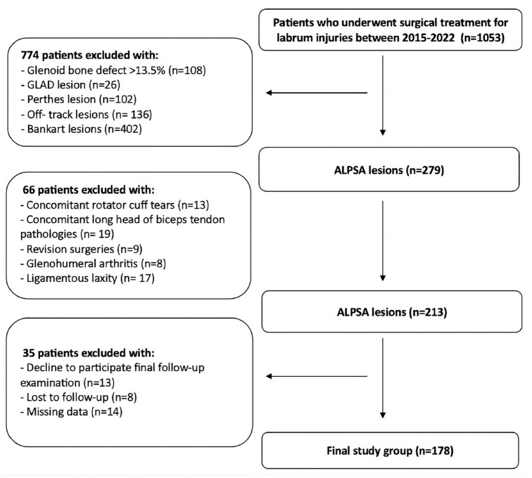
{"title":"Effect of ALPSA Tear Morphology on Redislocation Risk After Arthroscopic Repair.","authors":"Toygun Kagan Eren, Ethem Burak Oklaz, Ahmet Emin Okutan, Baran Sarikaya, Furkan Aral, Asim Ahmadov, Ulunay Kanatli","doi":"10.1177/23259671251360365","DOIUrl":null,"url":null,"abstract":"<p><strong>Background: </strong>Recent studies have emphasized the importance of lesion location and tear structure for understanding Bankart lesions; however, knowledge on anterior labroligamentous periosteal sleeve avulsion (ALPSA) lesion characteristics in anterior shoulder instability remains limited.</p><p><strong>Purpose: </strong>To evaluate the prevalence of various ALPSA lesion patterns and their effect on redislocation rates after labrum repair.</p><p><strong>Study design: </strong>Case-control study; Level of evidence, 3.</p><p><strong>Methods: </strong>Patients with ALPSA lesions who underwent arthroscopic labrum repair between 2015 and 2022 were retrospectively evaluated. Labrum tears were categorized into specific positions: isolated ALPSA lesions (3- to 5-o'clock position), ALPSA lesions with tears extending to the 1 o'clock position (1- to 5-o'clock position), and ALPSA lesions with tears extending into other positions. In addition, transverse tears that disrupted the circular continuity of the labrum were defined as radial tears. Patients were categorized as having no dislocated lesions or having redislocated lesions based on postoperative redislocation history. Descriptive data, tear extensions, radial tears, and patient-reported outcome measures (PROMs) were compared between the 2 groups.</p><p><strong>Results: </strong>The study included 178 patients (mean age, 25.7 ± 7.1 years), with a mean follow-up of 69.4 ± 27.2 months. Of these patients, 35 experienced lesion redislocation, while 143 patients did not experience lesion dislocation. In patients with no lesion dislocation, 43% of lesions were located in the 1- to 5-o'clock position, 36% in the 3- to 5-o'clock position, and 21% in other locations; in patients with lesion redislocation, 60% of lesions were observed in the 3- to 5-o'clock position, 29% in the 1- to 5-o'clock position, and 11% in other locations (<i>P</i> <sub>1-5</sub> = .04, <i>P</i> <sub>3-5</sub> = .001, and <i>P</i> <sub>others</sub> = .08). Radial tears were more frequent in the group with lesion redislocation (49%) compared with the group with no lesion dislocation (23%) (<i>P</i> < .001). Regression analysis demonstrated that radial tears (odds ratio [OR], 4.67) and the 3- to 5-o'clock lesion position (OR, 3.65) were significantly associated with redislocation (<i>P</i> = .01, <i>P</i> = .03, respectively). Both groups demonstrated significant improvements in PROMs compared with the preoperative period (<i>P</i> < .001). However, final follow-up PROMs were significantly worse in the group with lesion redislocation (<i>P</i> < .001).</p><p><strong>Conclusion: </strong>The present study demonstrated that an isolated ALPSA lesion at the 3- to 5-o'clock position and the presence of radial tears were independent factors increasing the risk of redislocation after arthroscopic ALPSA repair.</p>","PeriodicalId":19646,"journal":{"name":"Orthopaedic Journal of Sports Medicine","volume":"13 8","pages":"23259671251360365"},"PeriodicalIF":2.5000,"publicationDate":"2025-08-18","publicationTypes":"Journal Article","fieldsOfStudy":null,"isOpenAccess":false,"openAccessPdf":"https://www.ncbi.nlm.nih.gov/pmc/articles/PMC12361845/pdf/","citationCount":"0","resultStr":null,"platform":"Semanticscholar","paperid":null,"PeriodicalName":"Orthopaedic Journal of Sports Medicine","FirstCategoryId":"3","ListUrlMain":"https://doi.org/10.1177/23259671251360365","RegionNum":3,"RegionCategory":"医学","ArticlePicture":[],"TitleCN":null,"AbstractTextCN":null,"PMCID":null,"EPubDate":"2025/8/1 0:00:00","PubModel":"eCollection","JCR":"Q2","JCRName":"ORTHOPEDICS","Score":null,"Total":0}
引用次数: 0
Abstract
Background: Recent studies have emphasized the importance of lesion location and tear structure for understanding Bankart lesions; however, knowledge on anterior labroligamentous periosteal sleeve avulsion (ALPSA) lesion characteristics in anterior shoulder instability remains limited.
Purpose: To evaluate the prevalence of various ALPSA lesion patterns and their effect on redislocation rates after labrum repair.
Study design: Case-control study; Level of evidence, 3.
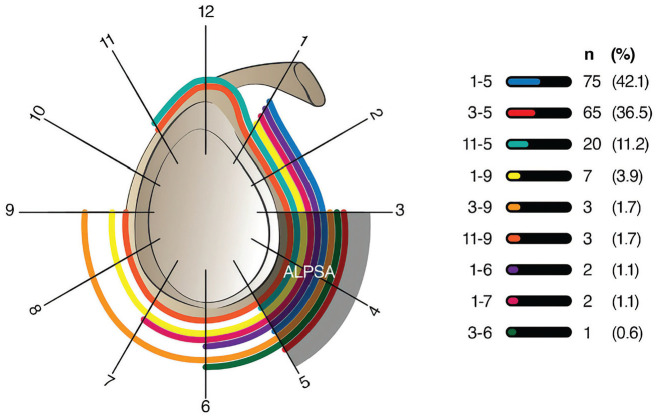
Methods: Patients with ALPSA lesions who underwent arthroscopic labrum repair between 2015 and 2022 were retrospectively evaluated. Labrum tears were categorized into specific positions: isolated ALPSA lesions (3- to 5-o'clock position), ALPSA lesions with tears extending to the 1 o'clock position (1- to 5-o'clock position), and ALPSA lesions with tears extending into other positions. In addition, transverse tears that disrupted the circular continuity of the labrum were defined as radial tears. Patients were categorized as having no dislocated lesions or having redislocated lesions based on postoperative redislocation history. Descriptive data, tear extensions, radial tears, and patient-reported outcome measures (PROMs) were compared between the 2 groups.
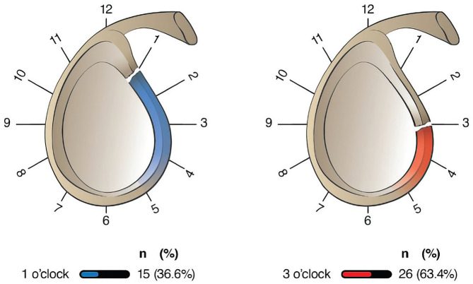
Results: The study included 178 patients (mean age, 25.7 ± 7.1 years), with a mean follow-up of 69.4 ± 27.2 months. Of these patients, 35 experienced lesion redislocation, while 143 patients did not experience lesion dislocation. In patients with no lesion dislocation, 43% of lesions were located in the 1- to 5-o'clock position, 36% in the 3- to 5-o'clock position, and 21% in other locations; in patients with lesion redislocation, 60% of lesions were observed in the 3- to 5-o'clock position, 29% in the 1- to 5-o'clock position, and 11% in other locations (P1-5 = .04, P3-5 = .001, and Pothers = .08). Radial tears were more frequent in the group with lesion redislocation (49%) compared with the group with no lesion dislocation (23%) (P < .001). Regression analysis demonstrated that radial tears (odds ratio [OR], 4.67) and the 3- to 5-o'clock lesion position (OR, 3.65) were significantly associated with redislocation (P = .01, P = .03, respectively). Both groups demonstrated significant improvements in PROMs compared with the preoperative period (P < .001). However, final follow-up PROMs were significantly worse in the group with lesion redislocation (P < .001).
Conclusion: The present study demonstrated that an isolated ALPSA lesion at the 3- to 5-o'clock position and the presence of radial tears were independent factors increasing the risk of redislocation after arthroscopic ALPSA repair.
期刊介绍:
The Orthopaedic Journal of Sports Medicine (OJSM), developed by the American Orthopaedic Society for Sports Medicine (AOSSM), is a global, peer-reviewed, open access journal that combines the interests of researchers and clinical practitioners across orthopaedic sports medicine, arthroscopy, and knee arthroplasty.
Topics include original research in the areas of:
-Orthopaedic Sports Medicine, including surgical and nonsurgical treatment of orthopaedic sports injuries
-Arthroscopic Surgery (Shoulder/Elbow/Wrist/Hip/Knee/Ankle/Foot)
-Relevant translational research
-Sports traumatology/epidemiology
-Knee and shoulder arthroplasty
The OJSM also publishes relevant systematic reviews and meta-analyses.
This journal is a member of the Committee on Publication Ethics (COPE).

 求助内容:
求助内容: 应助结果提醒方式:


