Taku Ebata, Koji Iwasaki, Tomoya Sato, Yoshiaki Hosokawa, Masanari Hamasaki, Dai Sato, Masatake Matsuoka, Tomohiro Onodera, Eiji Kondo, Norimasa Iwasaki
{"title":"The Relationship of Meniscal Extrusion to Subchondral Bone Density in Medial Knee Osteoarthritis.","authors":"Taku Ebata, Koji Iwasaki, Tomoya Sato, Yoshiaki Hosokawa, Masanari Hamasaki, Dai Sato, Masatake Matsuoka, Tomohiro Onodera, Eiji Kondo, Norimasa Iwasaki","doi":"10.1177/23259671251366424","DOIUrl":null,"url":null,"abstract":"<p><strong>Background: </strong>The hoop function of the meniscus plays a crucial role in stress distribution across the knee joint. While medial meniscal extrusion is known to contribute to the progression of medial knee osteoarthritis (OA) by altering load distribution within the knee joint, its exact effect on living humans remains unclear.</p><p><strong>Purpose: </strong>To investigate the influence of meniscal extrusion on subchondral bone density distribution in patients with medial knee OA.</p><p><strong>Study design: </strong>Cross-sectional study; Level of evidence, 3.</p><p><strong>Methods: </strong>This retrospective study included 59 patients with medial knee OA (OA group) and 19 control participants (non-OA group). Radiographic parameters, including the hip-knee-ankle angle (HKA) and meniscal extrusion ratio (MER), were assessed. The subchondral bone density was evaluated using computed tomography-osteoabsorptiometry to analyze the high-density area (HDA) in the medial and lateral compartments on the articular surface of the proximal tibia. Correlations between these parameters were assessed using single and multiple regression analyses, with subgroup analysis conducted in OA patients with and without meniscal tears.</p><p><strong>Results: </strong>In the OA group, the HKA, medial MER (MMER), and the ratio of the medial compartment HDA to the total HDA (medial ratio) were -7.4°, 64.8%, and 81.8%, respectively. In the non-OA group, these values were -2.1°, 12.5%, and 62.0%. Simple regression analysis showed that, in the OA group, the medial ratio was correlated with HKA (<i>R</i> <sup>2</sup> = 0.216; <i>P</i> < .001) and MMER (<i>R</i> <sup>2</sup> = 0.307; <i>P</i> < .001). Among non-OA participants, only MMER was correlated with the medial ratio (<i>R</i> <sup>2</sup> = 0.217; <i>P</i> = .045). The multivariable regression analysis demonstrated an adjusted <i>R</i> <sup>2</sup> value of 0.38 (<i>P</i> < .001) in the OA group. The standardized coefficients were 0.465 for MMER and -0.340 for HKA. Subgroup analysis further indicated that meniscal injury in OA patients amplified the effect of extrusion on subchondral bone density distribution, with an adjusted <i>R</i> <sup>2</sup> of 0.54 in the meniscal tear group.</p><p><strong>Conclusion: </strong>MMER had a greater influence on the mediolateral distribution of subchondral bone density in patients with medial knee OA than lower limb alignment, suggesting that the hoop function of the meniscus plays a more important role in altering stress distribution than leg alignment.</p>","PeriodicalId":19646,"journal":{"name":"Orthopaedic Journal of Sports Medicine","volume":"13 8","pages":"23259671251366424"},"PeriodicalIF":2.5000,"publicationDate":"2025-08-28","publicationTypes":"Journal Article","fieldsOfStudy":null,"isOpenAccess":false,"openAccessPdf":"https://www.ncbi.nlm.nih.gov/pmc/articles/PMC12394850/pdf/","citationCount":"0","resultStr":null,"platform":"Semanticscholar","paperid":null,"PeriodicalName":"Orthopaedic Journal of Sports Medicine","FirstCategoryId":"3","ListUrlMain":"https://doi.org/10.1177/23259671251366424","RegionNum":3,"RegionCategory":"医学","ArticlePicture":[],"TitleCN":null,"AbstractTextCN":null,"PMCID":null,"EPubDate":"2025/8/1 0:00:00","PubModel":"eCollection","JCR":"Q2","JCRName":"ORTHOPEDICS","Score":null,"Total":0}
引用次数: 0
Abstract
Background: The hoop function of the meniscus plays a crucial role in stress distribution across the knee joint. While medial meniscal extrusion is known to contribute to the progression of medial knee osteoarthritis (OA) by altering load distribution within the knee joint, its exact effect on living humans remains unclear.
Purpose: To investigate the influence of meniscal extrusion on subchondral bone density distribution in patients with medial knee OA.
Study design: Cross-sectional study; Level of evidence, 3.
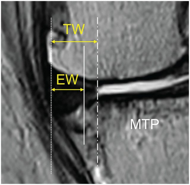
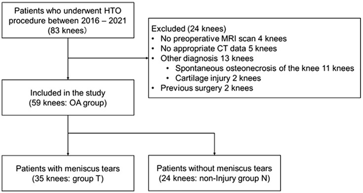
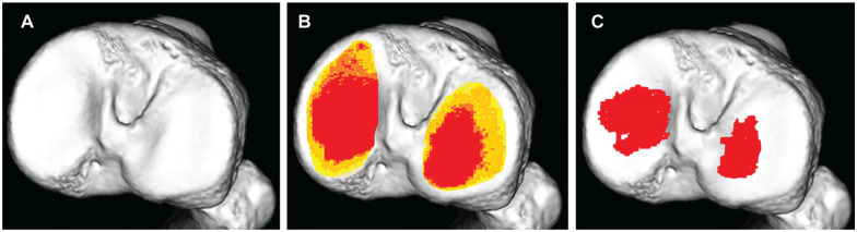
Methods: This retrospective study included 59 patients with medial knee OA (OA group) and 19 control participants (non-OA group). Radiographic parameters, including the hip-knee-ankle angle (HKA) and meniscal extrusion ratio (MER), were assessed. The subchondral bone density was evaluated using computed tomography-osteoabsorptiometry to analyze the high-density area (HDA) in the medial and lateral compartments on the articular surface of the proximal tibia. Correlations between these parameters were assessed using single and multiple regression analyses, with subgroup analysis conducted in OA patients with and without meniscal tears.
Results: In the OA group, the HKA, medial MER (MMER), and the ratio of the medial compartment HDA to the total HDA (medial ratio) were -7.4°, 64.8%, and 81.8%, respectively. In the non-OA group, these values were -2.1°, 12.5%, and 62.0%. Simple regression analysis showed that, in the OA group, the medial ratio was correlated with HKA (R2 = 0.216; P < .001) and MMER (R2 = 0.307; P < .001). Among non-OA participants, only MMER was correlated with the medial ratio (R2 = 0.217; P = .045). The multivariable regression analysis demonstrated an adjusted R2 value of 0.38 (P < .001) in the OA group. The standardized coefficients were 0.465 for MMER and -0.340 for HKA. Subgroup analysis further indicated that meniscal injury in OA patients amplified the effect of extrusion on subchondral bone density distribution, with an adjusted R2 of 0.54 in the meniscal tear group.
Conclusion: MMER had a greater influence on the mediolateral distribution of subchondral bone density in patients with medial knee OA than lower limb alignment, suggesting that the hoop function of the meniscus plays a more important role in altering stress distribution than leg alignment.
期刊介绍:
The Orthopaedic Journal of Sports Medicine (OJSM), developed by the American Orthopaedic Society for Sports Medicine (AOSSM), is a global, peer-reviewed, open access journal that combines the interests of researchers and clinical practitioners across orthopaedic sports medicine, arthroscopy, and knee arthroplasty.
Topics include original research in the areas of:
-Orthopaedic Sports Medicine, including surgical and nonsurgical treatment of orthopaedic sports injuries
-Arthroscopic Surgery (Shoulder/Elbow/Wrist/Hip/Knee/Ankle/Foot)
-Relevant translational research
-Sports traumatology/epidemiology
-Knee and shoulder arthroplasty
The OJSM also publishes relevant systematic reviews and meta-analyses.
This journal is a member of the Committee on Publication Ethics (COPE).

 求助内容:
求助内容: 应助结果提醒方式:


