{"title":"A Web-Deployed, Explainable AI System for Comprehensive Brain Tumor Diagnosis.","authors":"Serra Aksoy, Pinar Demircioglu, Ismail Bogrekci","doi":"10.3390/neurolint17080121","DOIUrl":null,"url":null,"abstract":"<p><strong>Background/objectives: </strong>Accurate diagnosis of brain tumors is one of the most important challenges in neuro-oncology since tumor classification and volumetric segmentation inform treatment planning. Two-dimensional classification and three-dimensional segmentation deep learning models can augment radiological workflows, particularly if paired with explainable AI techniques to improve model interpretability. The objective of this research was to develop a web-based brain tumor segmentation and classification diagnosis platform.</p><p><strong>Methods: </strong>A diagnosis system was developed combining 2D tumor classification and 3D volumetric segmentation. Classification employed a fine-tuned MobileNetV2 model trained on a glioma, meningioma, pituitary tumor, and normal control dataset. Segmentation employed a SegResNet model trained on BraTS multi-channel MRI with synthetic no-tumor data. A meta-classifier MLP was used for binary tumor detection from volumetric features. Explainability was offered using XRAI maps for 2D predictions and Gaussian overlays for 3D visualizations. The platform was incorporated into a web interface for clinical use.</p><p><strong>Results: </strong>MobileNetV2 2D model recorded 98.09% classification accuracy for tumor classification. 3D SegResNet obtained Dice coefficients around 68-70% for tumor segmentations. The MLP-based tumor detection module recorded 100% detection accuracy. Explainability modules could identify the area of the tumor, and saliency and overlay maps were consistent with real pathological features in both 2D and 3D.</p><p><strong>Conclusions: </strong>Deep learning diagnosis system possesses improved brain tumor classification and segmentation with interpretable outcomes by utilizing XAI techniques. Deployment as a web tool and a user-friendly interface made it suitable for clinical usage in radiology workflows.</p>","PeriodicalId":19130,"journal":{"name":"Neurology International","volume":"17 8","pages":""},"PeriodicalIF":3.0000,"publicationDate":"2025-08-04","publicationTypes":"Journal Article","fieldsOfStudy":null,"isOpenAccess":false,"openAccessPdf":"https://www.ncbi.nlm.nih.gov/pmc/articles/PMC12388561/pdf/","citationCount":"0","resultStr":null,"platform":"Semanticscholar","paperid":null,"PeriodicalName":"Neurology International","FirstCategoryId":"1085","ListUrlMain":"https://doi.org/10.3390/neurolint17080121","RegionNum":0,"RegionCategory":null,"ArticlePicture":[],"TitleCN":null,"AbstractTextCN":null,"PMCID":null,"EPubDate":"","PubModel":"","JCR":"Q2","JCRName":"CLINICAL NEUROLOGY","Score":null,"Total":0}
引用次数: 0
Abstract
Background/objectives: Accurate diagnosis of brain tumors is one of the most important challenges in neuro-oncology since tumor classification and volumetric segmentation inform treatment planning. Two-dimensional classification and three-dimensional segmentation deep learning models can augment radiological workflows, particularly if paired with explainable AI techniques to improve model interpretability. The objective of this research was to develop a web-based brain tumor segmentation and classification diagnosis platform.
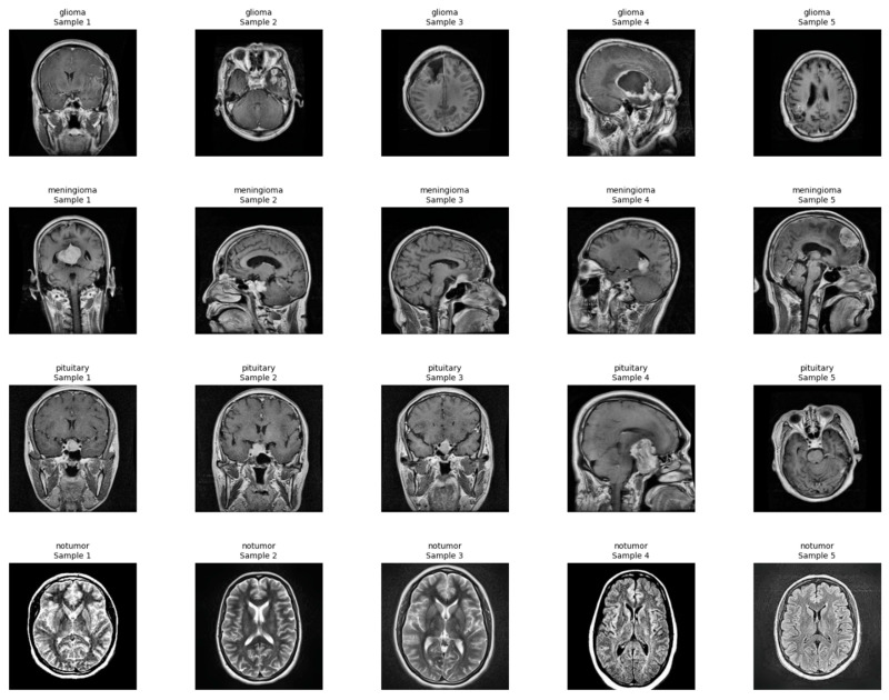
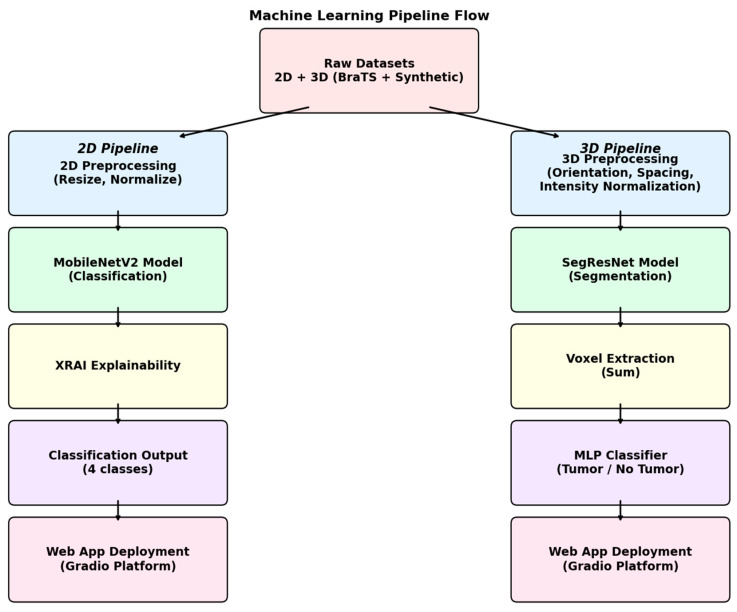
Methods: A diagnosis system was developed combining 2D tumor classification and 3D volumetric segmentation. Classification employed a fine-tuned MobileNetV2 model trained on a glioma, meningioma, pituitary tumor, and normal control dataset. Segmentation employed a SegResNet model trained on BraTS multi-channel MRI with synthetic no-tumor data. A meta-classifier MLP was used for binary tumor detection from volumetric features. Explainability was offered using XRAI maps for 2D predictions and Gaussian overlays for 3D visualizations. The platform was incorporated into a web interface for clinical use.
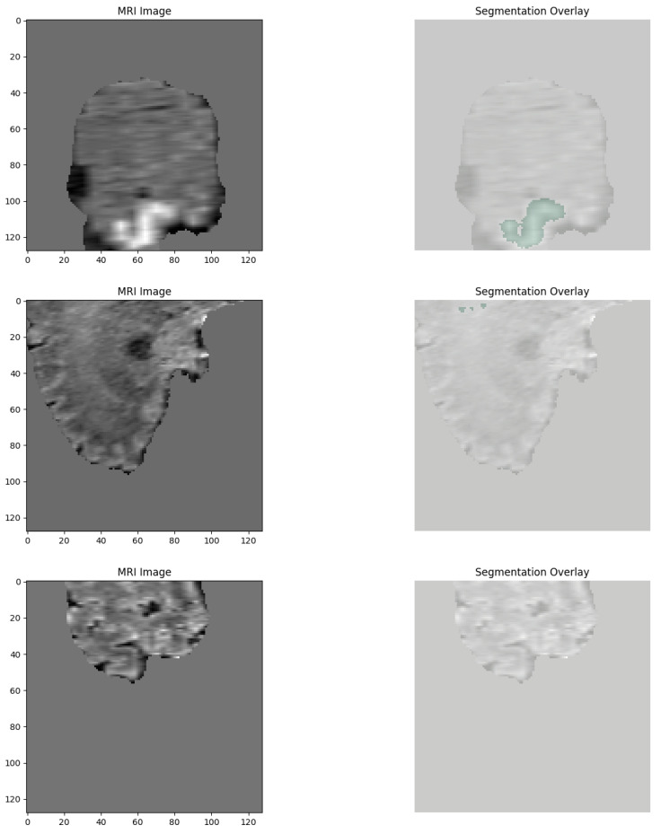
Results: MobileNetV2 2D model recorded 98.09% classification accuracy for tumor classification. 3D SegResNet obtained Dice coefficients around 68-70% for tumor segmentations. The MLP-based tumor detection module recorded 100% detection accuracy. Explainability modules could identify the area of the tumor, and saliency and overlay maps were consistent with real pathological features in both 2D and 3D.
Conclusions: Deep learning diagnosis system possesses improved brain tumor classification and segmentation with interpretable outcomes by utilizing XAI techniques. Deployment as a web tool and a user-friendly interface made it suitable for clinical usage in radiology workflows.

 求助内容:
求助内容: 应助结果提醒方式:


