{"title":"Three-Dimensional Magnetic Resonance Imaging of Median Nerve Volume for Assessing Carpal Tunnel Syndrome Severity.","authors":"Kazuki Hayakawa, Taku Suzuki, Katsuhiko Hayakawa, Yusuke Kawano, Takuji Iwamoto, Nobuyuki Fujita","doi":"10.1002/mus.70010","DOIUrl":null,"url":null,"abstract":"<p><strong>Introduction/aims: </strong>3D magnetic resonance imaging (MRI) is a tool for visualizing and quantifying the volume of the median nerve; however, the diagnostic value of the volume of the median nerve for assessing CTS severity is unclear. This study aimed to evaluate the utility of 3T MRI combined with three-dimensional (3D) imaging to assess the cross-sectional volume (CSV) of the median nerve for diagnosing carpal tunnel syndrome (CTS) and determining its severity.</p><p><strong>Methods: </strong>We used MRI to measure the CSV of the median nerve in 95 patients with CTS and 26 healthy controls. CTS severity was graded according to the Padua classification, and differences in CSV according to severity were analyzed.</p><p><strong>Results: </strong>The mean CSVs for the severity groups were as follows: control, 22.4 mm<sup>3</sup>; minimal, 28.8 mm<sup>3</sup>; moderate, 34.4 mm<sup>3</sup>; severe, 53.6 mm<sup>3</sup>; and extreme, 48.3 mm<sup>3</sup>. CSV increased with CTS severity, with significantly higher values in the severe and extreme groups compared with the moderate, minimal, and control groups.</p><p><strong>Discussion: </strong>This study showed that the CSV of the median nerve increased with disease severity, both visually and in three dimensions. This helps to visually understand the severity of CTS, and the association between morphology and disease severity may contribute to a better understanding of the pathophysiology of CTS.</p>","PeriodicalId":18968,"journal":{"name":"Muscle & Nerve","volume":" ","pages":"1117-1121"},"PeriodicalIF":3.1000,"publicationDate":"2025-11-01","publicationTypes":"Journal Article","fieldsOfStudy":null,"isOpenAccess":false,"openAccessPdf":"https://www.ncbi.nlm.nih.gov/pmc/articles/PMC12529045/pdf/","citationCount":"0","resultStr":null,"platform":"Semanticscholar","paperid":null,"PeriodicalName":"Muscle & Nerve","FirstCategoryId":"3","ListUrlMain":"https://doi.org/10.1002/mus.70010","RegionNum":3,"RegionCategory":"医学","ArticlePicture":[],"TitleCN":null,"AbstractTextCN":null,"PMCID":null,"EPubDate":"2025/8/28 0:00:00","PubModel":"Epub","JCR":"Q2","JCRName":"CLINICAL NEUROLOGY","Score":null,"Total":0}
引用次数: 0
Abstract
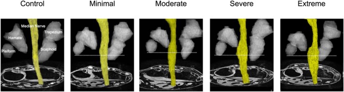
Introduction/aims: 3D magnetic resonance imaging (MRI) is a tool for visualizing and quantifying the volume of the median nerve; however, the diagnostic value of the volume of the median nerve for assessing CTS severity is unclear. This study aimed to evaluate the utility of 3T MRI combined with three-dimensional (3D) imaging to assess the cross-sectional volume (CSV) of the median nerve for diagnosing carpal tunnel syndrome (CTS) and determining its severity.
Methods: We used MRI to measure the CSV of the median nerve in 95 patients with CTS and 26 healthy controls. CTS severity was graded according to the Padua classification, and differences in CSV according to severity were analyzed.
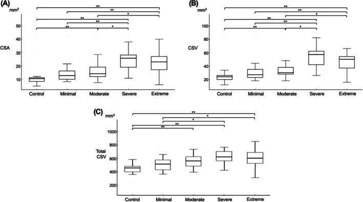
Results: The mean CSVs for the severity groups were as follows: control, 22.4 mm3; minimal, 28.8 mm3; moderate, 34.4 mm3; severe, 53.6 mm3; and extreme, 48.3 mm3. CSV increased with CTS severity, with significantly higher values in the severe and extreme groups compared with the moderate, minimal, and control groups.
Discussion: This study showed that the CSV of the median nerve increased with disease severity, both visually and in three dimensions. This helps to visually understand the severity of CTS, and the association between morphology and disease severity may contribute to a better understanding of the pathophysiology of CTS.
期刊介绍:
Muscle & Nerve is an international and interdisciplinary publication of original contributions, in both health and disease, concerning studies of the muscle, the neuromuscular junction, the peripheral motor, sensory and autonomic neurons, and the central nervous system where the behavior of the peripheral nervous system is clarified. Appearing monthly, Muscle & Nerve publishes clinical studies and clinically relevant research reports in the fields of anatomy, biochemistry, cell biology, electrophysiology and electrodiagnosis, epidemiology, genetics, immunology, pathology, pharmacology, physiology, toxicology, and virology. The Journal welcomes articles and reports on basic clinical electrophysiology and electrodiagnosis. We expedite some papers dealing with timely topics to keep up with the fast-moving pace of science, based on the referees'' recommendation.
 求助内容:
求助内容: 应助结果提醒方式:


