Diane Armao, Thomas W Bouldin, Diana X Bharucha-Goebel, Terry S Hartman, Steven J Gray, Rachel M Bailey, Dimah Saade, Joshua J Todd, Minal Jain, Melissa Waite, Carsten G Bönnemann, J Keith Smith
{"title":"T2 Hyperintensities in Gracile Tracts of Cervical Spinal Cord in Giant Axonal Neuropathy (GAN).","authors":"Diane Armao, Thomas W Bouldin, Diana X Bharucha-Goebel, Terry S Hartman, Steven J Gray, Rachel M Bailey, Dimah Saade, Joshua J Todd, Minal Jain, Melissa Waite, Carsten G Bönnemann, J Keith Smith","doi":"10.1002/mus.70004","DOIUrl":null,"url":null,"abstract":"<p><strong>Introduction/aims: </strong>Giant axonal neuropathy (GAN) is a hereditary neurodegenerative disease due to the absence or loss of function of the gigaxonin gene. Pathologic findings in GAN are those of \"dying-back\" axonal degeneration, in which the distal axon degenerates but the more proximal axon and neuronal cell body remain intact. Aims of this study were to (1) document imaging abnormalities that may occur in the spinal cords of GAN patients; and (2) assess histologically the spinal cords of GAN rodent models.</p><p><strong>Methods: </strong>A clinical trial of intrathecal (IT) scAAV9/JeT- GAN gene transfer provided a cohort of GAN patients for study. We examined spinal magnetic resonance imaging (MRI) studies from a subset of pretreatment GAN patients ages 6-14 years. For radiologic-pathologic correlation, we examined histologically spinal cords from GAN rodent models with pathological features of human GAN.</p><p><strong>Results: </strong>Of 10 GAN-patient spinal MRIs, 7 showed cervical or diffuse cord atrophy. Five MRIs additionally showed hyperintense, T2-signal abnormalities bilaterally in the cervical gracile tracts. Microscopy of GAN-rodent spinal cords revealed many actively degenerating axons in the cervical gracile tracts but few degenerating axons elsewhere in the cord.</p><p><strong>Discussion: </strong>The localization of spinal T-2 signal abnormalities to the cervical gracile tracts in GAN patients mirrors the localization of active dying-back axonal degeneration in GAN rodent models and suggests that these T2-signal abnormalities may be used as a surrogate marker of active axonal degeneration in the long tracts of the spinal cord in GAN and possibly other dying-back neurodegenerative diseases involving the spinal cord.</p>","PeriodicalId":18968,"journal":{"name":"Muscle & Nerve","volume":" ","pages":"1152-1155"},"PeriodicalIF":3.1000,"publicationDate":"2025-11-01","publicationTypes":"Journal Article","fieldsOfStudy":null,"isOpenAccess":false,"openAccessPdf":"https://www.ncbi.nlm.nih.gov/pmc/articles/PMC12529028/pdf/","citationCount":"0","resultStr":null,"platform":"Semanticscholar","paperid":null,"PeriodicalName":"Muscle & Nerve","FirstCategoryId":"3","ListUrlMain":"https://doi.org/10.1002/mus.70004","RegionNum":3,"RegionCategory":"医学","ArticlePicture":[],"TitleCN":null,"AbstractTextCN":null,"PMCID":null,"EPubDate":"2025/8/16 0:00:00","PubModel":"Epub","JCR":"Q2","JCRName":"CLINICAL NEUROLOGY","Score":null,"Total":0}
引用次数: 0
Abstract
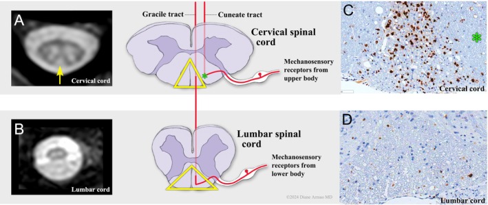
Introduction/aims: Giant axonal neuropathy (GAN) is a hereditary neurodegenerative disease due to the absence or loss of function of the gigaxonin gene. Pathologic findings in GAN are those of "dying-back" axonal degeneration, in which the distal axon degenerates but the more proximal axon and neuronal cell body remain intact. Aims of this study were to (1) document imaging abnormalities that may occur in the spinal cords of GAN patients; and (2) assess histologically the spinal cords of GAN rodent models.
Methods: A clinical trial of intrathecal (IT) scAAV9/JeT- GAN gene transfer provided a cohort of GAN patients for study. We examined spinal magnetic resonance imaging (MRI) studies from a subset of pretreatment GAN patients ages 6-14 years. For radiologic-pathologic correlation, we examined histologically spinal cords from GAN rodent models with pathological features of human GAN.
Results: Of 10 GAN-patient spinal MRIs, 7 showed cervical or diffuse cord atrophy. Five MRIs additionally showed hyperintense, T2-signal abnormalities bilaterally in the cervical gracile tracts. Microscopy of GAN-rodent spinal cords revealed many actively degenerating axons in the cervical gracile tracts but few degenerating axons elsewhere in the cord.
Discussion: The localization of spinal T-2 signal abnormalities to the cervical gracile tracts in GAN patients mirrors the localization of active dying-back axonal degeneration in GAN rodent models and suggests that these T2-signal abnormalities may be used as a surrogate marker of active axonal degeneration in the long tracts of the spinal cord in GAN and possibly other dying-back neurodegenerative diseases involving the spinal cord.
期刊介绍:
Muscle & Nerve is an international and interdisciplinary publication of original contributions, in both health and disease, concerning studies of the muscle, the neuromuscular junction, the peripheral motor, sensory and autonomic neurons, and the central nervous system where the behavior of the peripheral nervous system is clarified. Appearing monthly, Muscle & Nerve publishes clinical studies and clinically relevant research reports in the fields of anatomy, biochemistry, cell biology, electrophysiology and electrodiagnosis, epidemiology, genetics, immunology, pathology, pharmacology, physiology, toxicology, and virology. The Journal welcomes articles and reports on basic clinical electrophysiology and electrodiagnosis. We expedite some papers dealing with timely topics to keep up with the fast-moving pace of science, based on the referees'' recommendation.

 求助内容:
求助内容: 应助结果提醒方式:


