Olivia Blumberg, Quinton Wright, Ryan Breighner, Alexander Dash, Asher Lal, Zaina K Mosalam, Donald McMahon, Matthew F Koff, Jeri W Nieves, Eve Donnelly, Emily M Stein
{"title":"Bone texture by clinical magnetic resonance imaging is directly related to bone tissue maturity by Fourier-transform infrared spectroscopy.","authors":"Olivia Blumberg, Quinton Wright, Ryan Breighner, Alexander Dash, Asher Lal, Zaina K Mosalam, Donald McMahon, Matthew F Koff, Jeri W Nieves, Eve Donnelly, Emily M Stein","doi":"10.1093/jbmrpl/ziaf126","DOIUrl":null,"url":null,"abstract":"<p><p>Opportunistic screening for osteoporosis using images acquired for other purposes is a burgeoning area that may be of particular utility for the identification of surgical candidates with poor bone health. Texture analysis of clinical MRIs can be used to evaluate the heterogeneity of trabecular bone as a potential metric of bone quality. This cohort study investigated relationships between MRI-based vertebral trabecular bone texture and material properties by Fourier-transform infrared (FTIR) spectroscopy. We hypothesized that texture features from preoperative MRI images would reflect vertebral bone mineralization and collagen properties. In a cohort of 30 postmenopausal women (mean age 65) undergoing spine fusion surgery, T1-weighted MRI images were obtained using standard clinical sequences. A gray-level co-occurrence matrix was used to characterize the distribution and spatial organization of voxel intensities and derive texture features, including inverse difference moment, feature correlation, and contrast. Lumbar vertebral bone biopsies were obtained intraoperatively and analyzed with FTIR spectroscopy to assess composition, including metrics of mineral maturity (acid phosphate and carbonate:phosphate ratio). We found that vertebral trabecular bone texture by MRI was related to directly measured bone material properties: more heterogeneous texture was associated with less mature bone. Women with lower inverse difference moment had higher acid phosphate (<i>r</i> = -0.43, <i>p</i> < .02). Similarly, women with lower feature correlation had higher acid phosphate (<i>r</i> = -0.39, <i>p</i> < .04) and higher carbonate: phosphate (<i>r</i> = -0.47, <i>p</i> < .01). Women with higher contrast had higher acid phosphate (<i>r</i> = 0.381, <i>p</i> < .04). Our results suggest that preoperative MRI texture may predict intraoperative bone properties, specifically FTIR metrics of tissue age that may reflect local remodeling or microdamage repair processes. This finding supports the potential of MRI as a screening tool to identify individuals with abnormal bone quality.</p>","PeriodicalId":14611,"journal":{"name":"JBMR Plus","volume":"9 9","pages":"ziaf126"},"PeriodicalIF":2.4000,"publicationDate":"2025-07-29","publicationTypes":"Journal Article","fieldsOfStudy":null,"isOpenAccess":false,"openAccessPdf":"https://www.ncbi.nlm.nih.gov/pmc/articles/PMC12395337/pdf/","citationCount":"0","resultStr":null,"platform":"Semanticscholar","paperid":null,"PeriodicalName":"JBMR Plus","FirstCategoryId":"1085","ListUrlMain":"https://doi.org/10.1093/jbmrpl/ziaf126","RegionNum":0,"RegionCategory":null,"ArticlePicture":[],"TitleCN":null,"AbstractTextCN":null,"PMCID":null,"EPubDate":"2025/9/1 0:00:00","PubModel":"eCollection","JCR":"Q2","JCRName":"ENDOCRINOLOGY & METABOLISM","Score":null,"Total":0}
引用次数: 0
Abstract
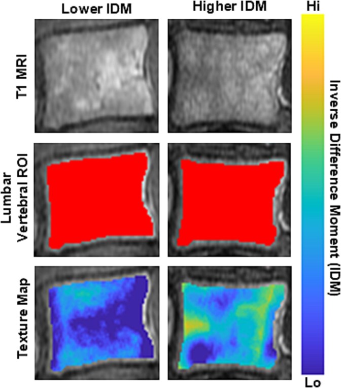
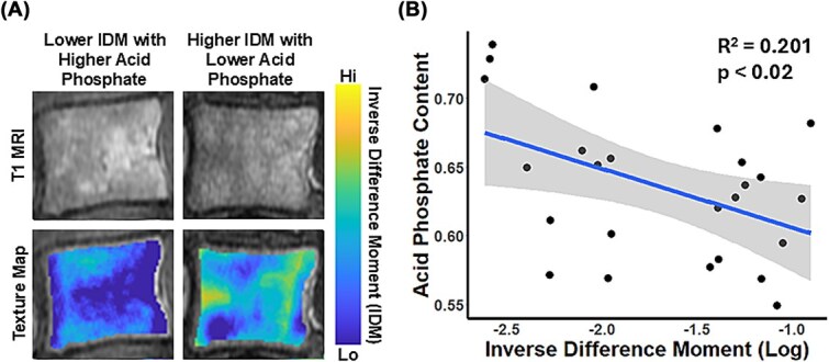
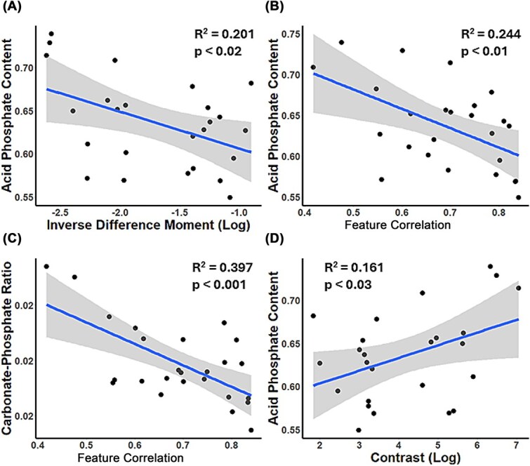
Opportunistic screening for osteoporosis using images acquired for other purposes is a burgeoning area that may be of particular utility for the identification of surgical candidates with poor bone health. Texture analysis of clinical MRIs can be used to evaluate the heterogeneity of trabecular bone as a potential metric of bone quality. This cohort study investigated relationships between MRI-based vertebral trabecular bone texture and material properties by Fourier-transform infrared (FTIR) spectroscopy. We hypothesized that texture features from preoperative MRI images would reflect vertebral bone mineralization and collagen properties. In a cohort of 30 postmenopausal women (mean age 65) undergoing spine fusion surgery, T1-weighted MRI images were obtained using standard clinical sequences. A gray-level co-occurrence matrix was used to characterize the distribution and spatial organization of voxel intensities and derive texture features, including inverse difference moment, feature correlation, and contrast. Lumbar vertebral bone biopsies were obtained intraoperatively and analyzed with FTIR spectroscopy to assess composition, including metrics of mineral maturity (acid phosphate and carbonate:phosphate ratio). We found that vertebral trabecular bone texture by MRI was related to directly measured bone material properties: more heterogeneous texture was associated with less mature bone. Women with lower inverse difference moment had higher acid phosphate (r = -0.43, p < .02). Similarly, women with lower feature correlation had higher acid phosphate (r = -0.39, p < .04) and higher carbonate: phosphate (r = -0.47, p < .01). Women with higher contrast had higher acid phosphate (r = 0.381, p < .04). Our results suggest that preoperative MRI texture may predict intraoperative bone properties, specifically FTIR metrics of tissue age that may reflect local remodeling or microdamage repair processes. This finding supports the potential of MRI as a screening tool to identify individuals with abnormal bone quality.
利用其他目的获取的图像对骨质疏松症进行机会性筛查是一个新兴的领域,可能对识别骨骼健康状况不佳的手术候选人特别有用。临床mri的纹理分析可用于评估骨小梁的异质性,作为骨质量的潜在指标。本队列研究通过傅里叶变换红外(FTIR)光谱研究了基于mri的椎小梁骨质地与材料特性之间的关系。我们假设术前MRI图像的纹理特征可以反映椎体骨矿化和胶原蛋白的特性。在30名接受脊柱融合手术的绝经后妇女(平均年龄65岁)队列中,使用标准临床序列获得t1加权MRI图像。利用灰度共现矩阵表征体素强度的分布和空间组织,导出纹理特征,包括逆差矩、特征相关性和对比度。术中获得腰椎骨活检,并用FTIR光谱分析评估成分,包括矿物成熟度指标(酸性磷酸盐和碳酸盐:磷酸盐比率)。我们发现MRI显示的椎小梁骨质地与直接测量的骨材料特性有关:质地越不均匀,骨成熟度越低。差负矩越小的女性酸性磷酸盐含量越高(r = -0.43, p r = -0.39, p r = -0.47, p r = 0.381, p

 求助内容:
求助内容: 应助结果提醒方式:


