{"title":"An MRI-based model for preoperative prediction of tertiary lymphoid structures in patients with gallbladder cancer.","authors":"Ying Xu, Zhuo Li, Weihua Zhi, Yi Yang, Jingzhong Ouyang, Yanzhao Zhou, Zeliang Ma, Sicong Wang, Lizhi Xie, Jianming Ying, Jinxue Zhou, Xinming Zhao, Feng Ye","doi":"10.1186/s13244-025-02007-4","DOIUrl":null,"url":null,"abstract":"<p><strong>Objectives: </strong>To predict tertiary lymphoid structures (TLSs) in gallbladder cancer (GBC) using preoperative magnetic resonance imaging (MRI)-based radiomics.</p><p><strong>Methods: </strong>Patients with GBC from two centres served as training (n = 129) and external validation (n = 44) cohorts. Radiomics features were extracted from six imaging sequences for inclusion in a radiomics model (Rad-score). Univariate and multivariate logistic regression were used to identify independent clinico-radiological predictors of TLS status. The clinical and radiomics models were integrated into a combined model. Areas under receiver operating characteristic curves (AUC) were used to assess model performance. The combined model was divided into low- and high-risk according to the cut-off value determined by the maximum Youden index of the ROC.</p><p><strong>Results: </strong>Intratumoural TLSs independently predicted RFS (p = 0.046). Eight features were included in the Rad-score. The clinical model included three independent predictors of TLS status (tumour height, liver invasion, and arterial-phase hypo-enhancement). In the training cohort, the combined model outperformed the separate clinical and radiomics models (AUC, 0.891 vs 0.870 and 0.775, respectively) and was externally valid. In both training and external cohorts, RFS in the low-risk group was substantially higher compared to the high-risk group. The low-risk group in the immunotherapy cohort had a significantly higher median overall survival than the high-risk group.</p><p><strong>Conclusions: </strong>The MRI-based combined model developed in this study can preoperatively predict intratumoural TLS status. It accurately stratified the RFS of patients after surgery and the OS of patients with immunotherapy.</p><p><strong>Critical relevance statement: </strong>This combined model is useful for predicting response and prognosis, not only for the recurrence-free survival of patients with GBC who have undergone surgery, but also for the overall survival of patients who have received immunotherapy KEY POINTS: Intratumoural TLSs independently predict recurrence-free survival of GBC. Our MRI-based combined model is a preoperative TLS marker. The combined model accurately stratifies postoperative/post-immunotherapy recurrence-free and overall survival of GBC.</p>","PeriodicalId":13639,"journal":{"name":"Insights into Imaging","volume":"16 1","pages":"189"},"PeriodicalIF":4.5000,"publicationDate":"2025-08-30","publicationTypes":"Journal Article","fieldsOfStudy":null,"isOpenAccess":false,"openAccessPdf":"https://www.ncbi.nlm.nih.gov/pmc/articles/PMC12398454/pdf/","citationCount":"0","resultStr":null,"platform":"Semanticscholar","paperid":null,"PeriodicalName":"Insights into Imaging","FirstCategoryId":"3","ListUrlMain":"https://doi.org/10.1186/s13244-025-02007-4","RegionNum":2,"RegionCategory":"医学","ArticlePicture":[],"TitleCN":null,"AbstractTextCN":null,"PMCID":null,"EPubDate":"","PubModel":"","JCR":"Q1","JCRName":"RADIOLOGY, NUCLEAR MEDICINE & MEDICAL IMAGING","Score":null,"Total":0}
引用次数: 0
Abstract
Objectives: To predict tertiary lymphoid structures (TLSs) in gallbladder cancer (GBC) using preoperative magnetic resonance imaging (MRI)-based radiomics.
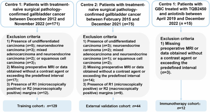
Methods: Patients with GBC from two centres served as training (n = 129) and external validation (n = 44) cohorts. Radiomics features were extracted from six imaging sequences for inclusion in a radiomics model (Rad-score). Univariate and multivariate logistic regression were used to identify independent clinico-radiological predictors of TLS status. The clinical and radiomics models were integrated into a combined model. Areas under receiver operating characteristic curves (AUC) were used to assess model performance. The combined model was divided into low- and high-risk according to the cut-off value determined by the maximum Youden index of the ROC.
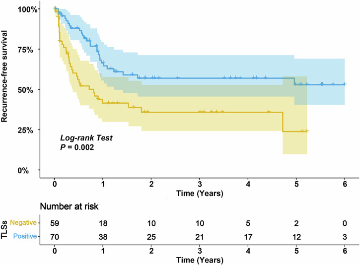
Results: Intratumoural TLSs independently predicted RFS (p = 0.046). Eight features were included in the Rad-score. The clinical model included three independent predictors of TLS status (tumour height, liver invasion, and arterial-phase hypo-enhancement). In the training cohort, the combined model outperformed the separate clinical and radiomics models (AUC, 0.891 vs 0.870 and 0.775, respectively) and was externally valid. In both training and external cohorts, RFS in the low-risk group was substantially higher compared to the high-risk group. The low-risk group in the immunotherapy cohort had a significantly higher median overall survival than the high-risk group.
Conclusions: The MRI-based combined model developed in this study can preoperatively predict intratumoural TLS status. It accurately stratified the RFS of patients after surgery and the OS of patients with immunotherapy.
Critical relevance statement: This combined model is useful for predicting response and prognosis, not only for the recurrence-free survival of patients with GBC who have undergone surgery, but also for the overall survival of patients who have received immunotherapy KEY POINTS: Intratumoural TLSs independently predict recurrence-free survival of GBC. Our MRI-based combined model is a preoperative TLS marker. The combined model accurately stratifies postoperative/post-immunotherapy recurrence-free and overall survival of GBC.
期刊介绍:
Insights into Imaging (I³) is a peer-reviewed open access journal published under the brand SpringerOpen. All content published in the journal is freely available online to anyone, anywhere!
I³ continuously updates scientific knowledge and progress in best-practice standards in radiology through the publication of original articles and state-of-the-art reviews and opinions, along with recommendations and statements from the leading radiological societies in Europe.
Founded by the European Society of Radiology (ESR), I³ creates a platform for educational material, guidelines and recommendations, and a forum for topics of controversy.
A balanced combination of review articles, original papers, short communications from European radiological congresses and information on society matters makes I³ an indispensable source for current information in this field.
I³ is owned by the ESR, however authors retain copyright to their article according to the Creative Commons Attribution License (see Copyright and License Agreement). All articles can be read, redistributed and reused for free, as long as the author of the original work is cited properly.
The open access fees (article-processing charges) for this journal are kindly sponsored by ESR for all Members.
The journal went open access in 2012, which means that all articles published since then are freely available online.

 求助内容:
求助内容: 应助结果提醒方式:


