Heda Melinda Nataprawira, Intan M W Dewi, I Gusti Agung Ayu Novi Wiraningrat, Citra Cesilia
{"title":"Imaging of post-tuberculosis lung disease cases in children and adolescent survivors: a systematic review.","authors":"Heda Melinda Nataprawira, Intan M W Dewi, I Gusti Agung Ayu Novi Wiraningrat, Citra Cesilia","doi":"10.1080/20018525.2025.2547515","DOIUrl":null,"url":null,"abstract":"<p><strong>Introduction: </strong>Post-tuberculosis lung disease (PTLD) causes health problems among pulmonary TB (PTB) survivors. Post-TB patients may suffer from chronic respiratory symptoms, declining lung function, and persistent radiological abnormalities. However, studies regarding PTLD in children and adolescents are still scarce. Patterns of radiological abnormalities, including chest X-ray (CXR) imaging, high-resolution computed tomography (HRCT), and magnetic resonance imaging (MRI) in post-TB children, and adolescents are not fully understood.</p><p><strong>Aim: </strong>In this study, we aim to review and analyse radiological features in children and adolescent TB survivors of the literature on the differences in imaging findings in drug-resistant (DR) and drug-sensitive tuberculosis (DS TB) children and adolescent TB survivors.</p><p><strong>Method: </strong>We performed a systematic review to determine imaging patterns of DR and DS TB in children and adolescent survivors. Data collected include study design, number of subjects, age, TB category, treatment duration, time of evaluation, and imaging patterns. We searched MEDLINE/Pubmed, Google Scholar, Science Direct, Wiley Online Library, Cochrane Library, and Proquest and included four studies for data analysis. Study quality was assessed using a modified Newcastle-Ottawa score.</p><p><strong>Result: </strong>Studies included 151 children and adolescents aged 0-17 years. Three out of four studies were conducted on DS-TB patients and one study compared DS- and DR-TB. Radiological abnormalities observed by CXR at TB treatment completion include calcification in the presence or absence of fibrosis, bronchiectasis, and destroyed lung, or lymphoid interstitial pneumonitis. Micronodules are most often seen in HRCT in the acute early stages of TB and were not seen in standard chest radiography. Cavities persisted in almost 50% of patients after TB treatment and fibrotic changes increased after treatment.</p><p><strong>Conclusion: </strong>Imaging abnormalities after TB treatment are often seen in children and adolescents. Imaging evaluation should be performed in PTB survivors, especially in those with moderate or advanced lesions during active disease and those with severe clinical manifestations.</p>","PeriodicalId":11872,"journal":{"name":"European Clinical Respiratory Journal","volume":"12 1","pages":"2547515"},"PeriodicalIF":1.4000,"publicationDate":"2025-08-18","publicationTypes":"Journal Article","fieldsOfStudy":null,"isOpenAccess":false,"openAccessPdf":"https://www.ncbi.nlm.nih.gov/pmc/articles/PMC12364111/pdf/","citationCount":"0","resultStr":null,"platform":"Semanticscholar","paperid":null,"PeriodicalName":"European Clinical Respiratory Journal","FirstCategoryId":"1085","ListUrlMain":"https://doi.org/10.1080/20018525.2025.2547515","RegionNum":0,"RegionCategory":null,"ArticlePicture":[],"TitleCN":null,"AbstractTextCN":null,"PMCID":null,"EPubDate":"2025/1/1 0:00:00","PubModel":"eCollection","JCR":"Q3","JCRName":"RESPIRATORY SYSTEM","Score":null,"Total":0}
引用次数: 0
Abstract
Introduction: Post-tuberculosis lung disease (PTLD) causes health problems among pulmonary TB (PTB) survivors. Post-TB patients may suffer from chronic respiratory symptoms, declining lung function, and persistent radiological abnormalities. However, studies regarding PTLD in children and adolescents are still scarce. Patterns of radiological abnormalities, including chest X-ray (CXR) imaging, high-resolution computed tomography (HRCT), and magnetic resonance imaging (MRI) in post-TB children, and adolescents are not fully understood.
Aim: In this study, we aim to review and analyse radiological features in children and adolescent TB survivors of the literature on the differences in imaging findings in drug-resistant (DR) and drug-sensitive tuberculosis (DS TB) children and adolescent TB survivors.
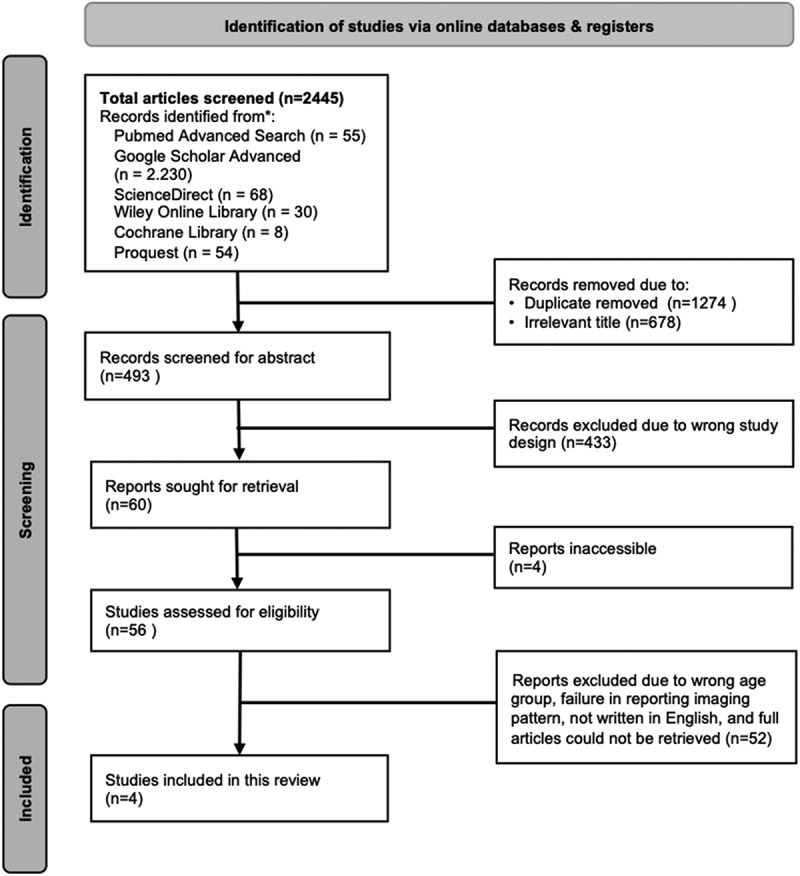
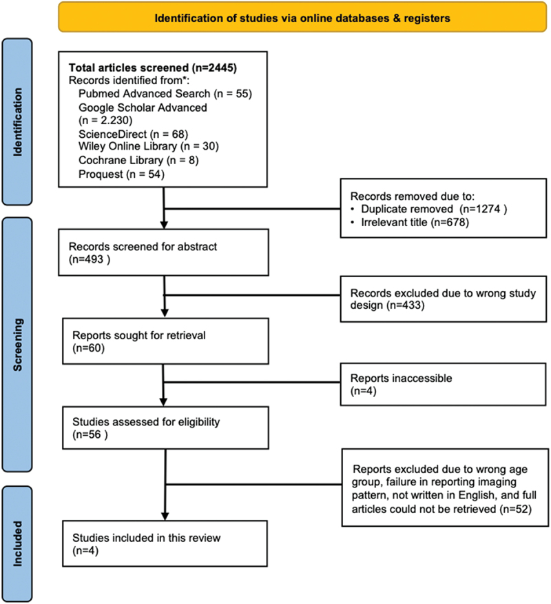
Method: We performed a systematic review to determine imaging patterns of DR and DS TB in children and adolescent survivors. Data collected include study design, number of subjects, age, TB category, treatment duration, time of evaluation, and imaging patterns. We searched MEDLINE/Pubmed, Google Scholar, Science Direct, Wiley Online Library, Cochrane Library, and Proquest and included four studies for data analysis. Study quality was assessed using a modified Newcastle-Ottawa score.
Result: Studies included 151 children and adolescents aged 0-17 years. Three out of four studies were conducted on DS-TB patients and one study compared DS- and DR-TB. Radiological abnormalities observed by CXR at TB treatment completion include calcification in the presence or absence of fibrosis, bronchiectasis, and destroyed lung, or lymphoid interstitial pneumonitis. Micronodules are most often seen in HRCT in the acute early stages of TB and were not seen in standard chest radiography. Cavities persisted in almost 50% of patients after TB treatment and fibrotic changes increased after treatment.
Conclusion: Imaging abnormalities after TB treatment are often seen in children and adolescents. Imaging evaluation should be performed in PTB survivors, especially in those with moderate or advanced lesions during active disease and those with severe clinical manifestations.
 求助内容:
求助内容: 应助结果提醒方式:


