Elin Trägårdh, Johannes Ulén, Olof Enqvist, Måns Larsson, Kristian Valind, David Minarik, Lars Edenbrandt
{"title":"A fully automated AI-based method for tumour detection and quantification on [<sup>18</sup>F]PSMA-1007 PET-CT images in prostate cancer.","authors":"Elin Trägårdh, Johannes Ulén, Olof Enqvist, Måns Larsson, Kristian Valind, David Minarik, Lars Edenbrandt","doi":"10.1186/s40658-025-00786-9","DOIUrl":null,"url":null,"abstract":"<p><strong>Background: </strong>In this study, we further developed an artificial intelligence (AI)-based method for the detection and quantification of tumours in the prostate, lymph nodes and bone in prostate-specific membrane antigen (PSMA)-targeting positron emission tomography with computed tomography (PET-CT) images.</p><p><strong>Methods: </strong>A total of 1064 [<sup>18</sup>F]PSMA-1007 PET-CT scans were used (approximately twice as many compared to our previous AI model), of which 120 were used as test set. Suspected lesions were manually annotated and used as ground truth. A convolutional neural network was developed and trained. The sensitivity and positive predictive value (PPV) were calculated using two sets of manual segmentations as reference. Results were also compared to our previously developed AI method. The correlation between manually and AI-based calculations of total lesion volume (TLV) and total lesion uptake (TLU) were calculated.</p><p><strong>Results: </strong>The sensitivities of the AI method were 85% for prostate tumour/recurrence, 91% for lymph node metastases and 61% for bone metastases (82%, 86% and 70% for manual readings and 66%, 88% and 71% for the old AI method). The PPVs of the AI method were 85%, 83% and 58%, respectively (63%, 86% and 39% for manual readings, and 69%, 70% and 39% for the old AI method). The correlations between manual and AI-based calculations of TLV and TLU ranged from r = 0.62 to r = 0.96.</p><p><strong>Conclusion: </strong>The performance of the newly developed and fully automated AI-based method for detecting and quantifying prostate tumour and suspected lymph node and bone metastases increased significantly, especially the PPV. The AI method is freely available to other researchers ( www.recomia.org ).</p>","PeriodicalId":11559,"journal":{"name":"EJNMMI Physics","volume":"12 1","pages":"78"},"PeriodicalIF":3.2000,"publicationDate":"2025-08-20","publicationTypes":"Journal Article","fieldsOfStudy":null,"isOpenAccess":false,"openAccessPdf":"https://www.ncbi.nlm.nih.gov/pmc/articles/PMC12367631/pdf/","citationCount":"0","resultStr":null,"platform":"Semanticscholar","paperid":null,"PeriodicalName":"EJNMMI Physics","FirstCategoryId":"3","ListUrlMain":"https://doi.org/10.1186/s40658-025-00786-9","RegionNum":2,"RegionCategory":"医学","ArticlePicture":[],"TitleCN":null,"AbstractTextCN":null,"PMCID":null,"EPubDate":"","PubModel":"","JCR":"Q2","JCRName":"RADIOLOGY, NUCLEAR MEDICINE & MEDICAL IMAGING","Score":null,"Total":0}
引用次数: 0
Abstract
Background: In this study, we further developed an artificial intelligence (AI)-based method for the detection and quantification of tumours in the prostate, lymph nodes and bone in prostate-specific membrane antigen (PSMA)-targeting positron emission tomography with computed tomography (PET-CT) images.
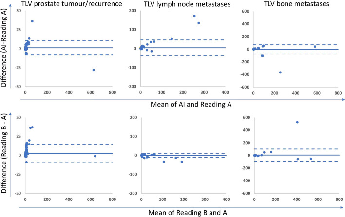
Methods: A total of 1064 [18F]PSMA-1007 PET-CT scans were used (approximately twice as many compared to our previous AI model), of which 120 were used as test set. Suspected lesions were manually annotated and used as ground truth. A convolutional neural network was developed and trained. The sensitivity and positive predictive value (PPV) were calculated using two sets of manual segmentations as reference. Results were also compared to our previously developed AI method. The correlation between manually and AI-based calculations of total lesion volume (TLV) and total lesion uptake (TLU) were calculated.
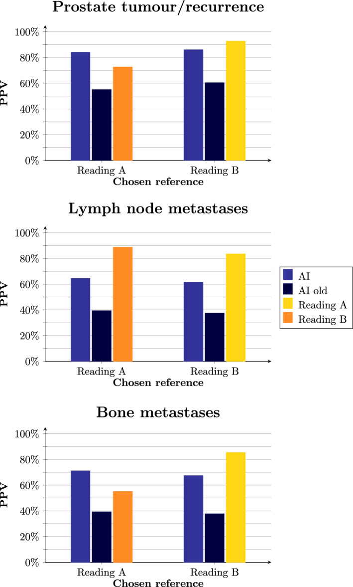
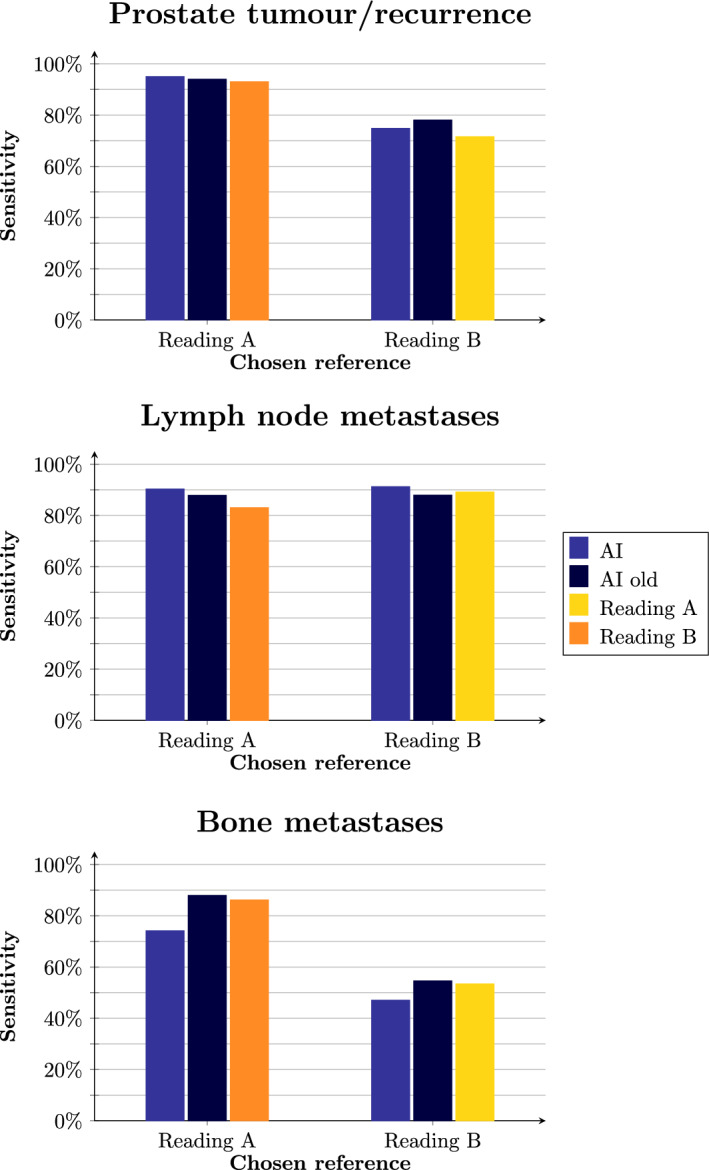
Results: The sensitivities of the AI method were 85% for prostate tumour/recurrence, 91% for lymph node metastases and 61% for bone metastases (82%, 86% and 70% for manual readings and 66%, 88% and 71% for the old AI method). The PPVs of the AI method were 85%, 83% and 58%, respectively (63%, 86% and 39% for manual readings, and 69%, 70% and 39% for the old AI method). The correlations between manual and AI-based calculations of TLV and TLU ranged from r = 0.62 to r = 0.96.
Conclusion: The performance of the newly developed and fully automated AI-based method for detecting and quantifying prostate tumour and suspected lymph node and bone metastases increased significantly, especially the PPV. The AI method is freely available to other researchers ( www.recomia.org ).
期刊介绍:
EJNMMI Physics is an international platform for scientists, users and adopters of nuclear medicine with a particular interest in physics matters. As a companion journal to the European Journal of Nuclear Medicine and Molecular Imaging, this journal has a multi-disciplinary approach and welcomes original materials and studies with a focus on applied physics and mathematics as well as imaging systems engineering and prototyping in nuclear medicine. This includes physics-driven approaches or algorithms supported by physics that foster early clinical adoption of nuclear medicine imaging and therapy.

 求助内容:
求助内容: 应助结果提醒方式:


