Development of a multi-feature predictive model for risk stratification in stage IB-IIA non-small cell lung cancer: a multicenter analysis
IF 3.3
3区 医学
Q1 RADIOLOGY, NUCLEAR MEDICINE & MEDICAL IMAGING
引用次数: 0
Abstract
Objective
This study aimed to develop a comprehensive risk stratification model for stage IB-IIA non-small cell lung cancer (NSCLC) by integrating clinicopathological data with pre-treatment CT imaging.
Methods
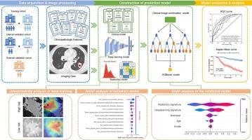
This retrospective study included three independent cohorts of patients with stage IB-IIA NSCLC for model development and validation (Training: n = 370; Internal validation: n = 120; External validation: n = 70). Disease-free survival (DFS) was the primary endpoint. Radiomics features were extracted from both tumoral and peritumoral regions of CT images to construct a radiomics model. A ResNet50-based deep learning architecture was adopted to develop a deep learning model using CT imaging data. Logistic regression was used to identify significant clinicopathological factors. These components were integrated into a multi-feature combined model (CRD model) that utilized clinicopathological, radiomics, and deep learning features for DFS prediction. Model interpretability was assessed using the SHapley Additive exPlanations (SHAP) method.
Results
The combined CRD model demonstrated superior performance in predicting DFS, achieving areas under the curve (AUC) of 0.865, 0.798, and 0.803 in the training, internal validation, and external validation cohorts, respectively. Patients were stratified into high- and low-risk groups using the CRD model, and in the external validation cohort, the hazard ratio (HR) for high-risk patients was 17.509, with a C-index of 0.73. SHAP analysis revealed that radiomics features contributed most significantly to the performance of the CRD model.
Conclusions
The multi-feature combined model effectively predicts DFS and identifies high-risk patients with stage IB-IIA NSCLC. It could facilitate personalized postoperative treatment strategies, improving patient outcomes.
IB-IIA期非小细胞肺癌多特征风险分层预测模型的建立:一项多中心分析
目的将临床病理资料与治疗前CT影像相结合,建立IB-IIA期非小细胞肺癌(NSCLC)的综合风险分层模型。方法本回顾性研究纳入三个独立的IB-IIA期非小细胞肺癌患者队列,用于模型开发和验证(训练组:n = 370;内部验证:n = 120;外部验证:n = 70)。无病生存期(DFS)是主要终点。从CT图像的肿瘤和肿瘤周围区域提取放射组学特征,构建放射组学模型。采用基于resnet50的深度学习架构,利用CT成像数据建立深度学习模型。采用Logistic回归分析确定显著的临床病理因素。这些成分被整合到一个多特征组合模型(CRD模型)中,该模型利用临床病理学、放射组学和深度学习特征进行DFS预测。采用SHapley加性解释(SHAP)方法评估模型可解释性。结果联合CRD模型对DFS的预测效果较好,在训练组、内部验证组和外部验证组的曲线下面积(AUC)分别为0.865、0.798和0.803。采用CRD模型将患者分为高危组和低危组,在外部验证队列中,高危患者的风险比(HR)为17.509,c指数为0.73。SHAP分析显示放射组学特征对CRD模型的性能贡献最大。结论多特征联合模型可有效预测DFS,识别IB-IIA期NSCLC高危患者。它可以促进个性化的术后治疗策略,改善患者的预后。
本文章由计算机程序翻译,如有差异,请以英文原文为准。
求助全文
约1分钟内获得全文
求助全文
来源期刊

CiteScore
6.70
自引率
3.00%
发文量
398
审稿时长
42 days
期刊介绍:
European Journal of Radiology is an international journal which aims to communicate to its readers, state-of-the-art information on imaging developments in the form of high quality original research articles and timely reviews on current developments in the field.
Its audience includes clinicians at all levels of training including radiology trainees, newly qualified imaging specialists and the experienced radiologist. Its aim is to inform efficient, appropriate and evidence-based imaging practice to the benefit of patients worldwide.
 求助内容:
求助内容: 应助结果提醒方式:


