Advances in neuroimaging applications of quantitative susceptibility mapping
引用次数: 0
Abstract
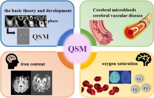
This review article delves into the advancements of quantitative susceptibility mapping (QSM) in neuroimaging, highlighting its utility in detecting and quantifying magnetic susceptibility differences in tissues, particularly for paramagnetic substances like iron and diamagnetic substances such as calcifications in the brain. QSM has revolutionized the diagnosis and monitoring of neurodegenerative diseases by enabling the precise measurement of brain iron deposition and blood oxygen saturation. The review is partitioned into three sections. The first section underscores QSM's role in clinical applications related to microhemorrhages, cerebral amyloidosis, intracranial hematomas, and cerebrovascular malformations. The second section focuses on QSM's application in mapping iron content in neurodegenerative diseases such as Parkinson's disease and Alzheimer's disease. The final section discusses QSM's potential in assessing stroke by measuring oxygen saturation. The article also outlines the basic theory and development of QSM, emphasizing the importance of echo time selection for accurate QSM results. Challenges in clinical applications and future directions, including the integration of AI technology for image reconstruction and data analysis, are also discussed. QSM's ability to differentiate between microbleeds and calcifications, assess dynamic susceptibility changes in intracranial hematomas, and guide thrombolytic strategies in acute cerebrovascular disease is highlighted. The review concludes by emphasizing the need for further optimization of QSM algorithms and the expansion of its applications in biomedical imaging.
定量易感图谱在神经影像学中的应用进展
本文综述了定量易感性制图(QSM)在神经影像学中的研究进展,重点介绍了其在检测和定量组织磁化率差异方面的应用,特别是对铁等顺磁性物质和脑钙化等反磁性物质。QSM通过精确测量脑铁沉积和血氧饱和度,彻底改变了神经退行性疾病的诊断和监测。这篇综述分为三个部分。第一部分强调了QSM在与微出血、脑淀粉样变性、颅内血肿和脑血管畸形相关的临床应用中的作用。第二部分重点介绍QSM在帕金森病和阿尔茨海默病等神经退行性疾病中铁含量制图中的应用。最后一节讨论了通过测量血氧饱和度来评估脑卒中的QSM的潜力。本文还概述了QSM的基本理论和发展,强调了回波时间选择对准确的QSM结果的重要性。还讨论了临床应用中的挑战和未来方向,包括将人工智能技术集成到图像重建和数据分析中。QSM能够区分微出血和钙化,评估颅内血肿的动态易感性变化,并指导急性脑血管疾病的溶栓策略。最后强调了QSM算法的进一步优化和在生物医学成像中的应用。
本文章由计算机程序翻译,如有差异,请以英文原文为准。
求助全文
约1分钟内获得全文
求助全文

 求助内容:
求助内容: 应助结果提醒方式:


