Optimizing spinal cord lesion segmentation using hierarchical classification and U-NET based segmentation model
IF 4.5
Q2 COMPUTER SCIENCE, THEORY & METHODS
引用次数: 0
Abstract
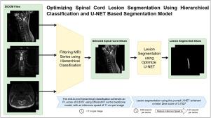
Rapid and accurate identification of spinal cord lesions is crucial in emergency medical care; however, current automated analysis systems face significant challenges in terms of processing speed and accuracy. This study presents a novel approach that combines hierarchical classification with segmentation models for automated spinal cord lesion analysis that can operate without human intervention, from image selection to lesion segmentation. We developed and evaluated a multistage classification system utilizing state-of-the-art efficient vision models, including EfficientVit, MobileNetV4, and RepVit, and compared it with a ResNet baseline. We incorporated a transfer learning strategy for lesion segmentation, leveraging pre-trained models to improve lesion boundary delineation. In addition, we introduced a synthetic lesion augmentation framework to address data scarcity and improve the generalization of the model. The system was trained and validated using six public datasets, including RadImageNet (1.4M images), and two private hospital datasets comprising 351 patients with spinal MRI scans. Our hierarchical classification approach achieved an end-to-end F1 score of 0.8357 using EfficientVit, whereas our optimized U-Net segmentation model attained a mean Dice score of 0.7527, surpassing previous approaches. Notably, the system achieved a ninefold increase in processing speed, reducing the lesion segmentation analysis time from 9.86 to 1.04 s per series and achieving a classification inference speed of 11.47 ms per image. Our findings suggest that this approach may support efficient automated spinal cord lesion analysis in resource-constrained clinical settings, potentially aiding diagnosis in emergency care, supporting radiologist workflow, and increasing the accessibility of spinal cord lesion analysis.
基于层次分类和U-NET分割模型的脊髓损伤分割优化
快速准确地识别脊髓病变在紧急医疗护理中至关重要;然而,当前的自动化分析系统在处理速度和准确性方面面临着重大挑战。本研究提出了一种新的方法,将分层分类与分割模型相结合,用于自动脊髓病变分析,从图像选择到病变分割,无需人工干预。我们利用最先进的高效视觉模型(包括EfficientVit、MobileNetV4和RepVit)开发并评估了一个多级分类系统,并将其与ResNet基线进行了比较。我们将转移学习策略用于病灶分割,利用预先训练的模型来改善病灶边界的描绘。此外,我们引入了一个综合损伤增强框架来解决数据稀缺问题,提高模型的泛化能力。该系统使用六个公共数据集(包括RadImageNet(140万张图像))和两个私立医院数据集(包括351名脊柱MRI扫描患者)进行训练和验证。我们的分层分类方法使用EfficientVit实现了端到端的F1得分为0.8357,而我们优化的U-Net分割模型的平均Dice得分为0.7527,超过了以前的方法。值得注意的是,该系统的处理速度提高了9倍,将病灶分割分析时间从9.86 s /序列降低到1.04 s /序列,实现了11.47 ms /图像的分类推理速度。我们的研究结果表明,这种方法可以在资源有限的临床环境中支持有效的脊髓病变自动分析,潜在地帮助急诊诊断,支持放射科医生的工作流程,并增加脊髓病变分析的可及性。
本文章由计算机程序翻译,如有差异,请以英文原文为准。
求助全文
约1分钟内获得全文
求助全文

 求助内容:
求助内容: 应助结果提醒方式:


