Automatic analysis of carpal angles using dynamic CT imaging: Reference values in healthy wrists and assessment of scapholunate ligament injuries
IF 3.3
3区 医学
Q1 RADIOLOGY, NUCLEAR MEDICINE & MEDICAL IMAGING
引用次数: 0
Abstract
Introduction
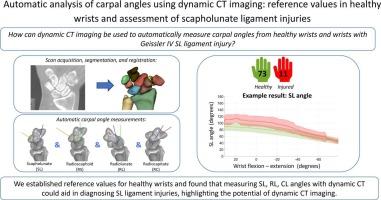
Dynamic CT imaging is a promising modality for evaluating wrist pathologies like scapholunate ligament (SL) injuries. The primary objective of this study is to extract carpal angles from dynamic CT datasets using an automated motion analysis algorithm to provide reference values for healthy wrist motion. Secondly, the feasibility of this automatic method to detect SL ligament pathology was evaluated.
Methods
Dynamic CT scans of healthy wrists and wrists with arthroscopically-confirmed complete SL injuries (Geissler IV) were analysed. Each scan consisted of one static image and two dynamic imaging sequences: wrist radial-ulnar deviation (RUD) and flexion–extension (FE). Bones were automatically segmented, and the radioscaphoid (RSA), scapholunate (SLA), capitolunate (CLA), and radiolunate (RLA) angles were automatically determined in each wrist position. A linear mixed model was applied to compare carpal angles between the two groups (p < 0.05).
Results
A total of 84 wrists scans were analysed, of which 73 healthy and 11 injured. Reference values for healthy wrists were provided, with an average and maximum 95% CI width during all movements of 5°and 7°, respectively. Feasibility analysis showed that the SLA, CLA, and RLA were different between the healthy and injured groups during all movements. No differences were found for the RSA.
Conclusion
Reference values of the moving wrist of healthy participants were automatically extracted. Furthermore, our results suggest that the RLA, CLA, and SLA may be useful parameters for distinguishing wrists with complete SL injuries from healthy ones, making the automatic approach feasible.
动态CT自动分析腕角:健康腕关节的参考值和舟月韧带损伤的评估
动态CT成像是评估腕关节病变如舟月骨韧带(SL)损伤的一种很有前途的方式。本研究的主要目的是使用自动运动分析算法从动态CT数据集中提取腕角,为健康的手腕运动提供参考值。其次,评估该方法自动检测骶髂韧带病理的可行性。方法对健康腕关节和经关节镜确认的完全性骶髂关节损伤(Geissler IV型)患者的动态CT扫描结果进行分析。每次扫描包括一个静态图像和两个动态成像序列:手腕桡尺偏差(RUD)和屈伸(FE)。骨自动分割,自动确定每个腕位的桡舟状骨(RSA)、舟月骨(SLA)、头月骨(CLA)和桡月骨(RLA)角度。采用线性混合模型比较两组患者腕角(p < 0.05)。结果共分析84例腕关节扫描,其中健康73例,受伤11例。提供了健康手腕的参考值,在所有运动中,95% CI宽度的平均值和最大值分别为5°和7°。可行性分析表明,在所有运动过程中,健康组和损伤组的SLA、CLA和RLA存在差异。RSA没有发现差异。结论自动提取了健康受试者运动腕关节的参考值。此外,我们的研究结果表明,RLA、CLA和SLA可能是区分完全SL损伤腕关节与健康腕关节的有用参数,使自动入路成为可能。
本文章由计算机程序翻译,如有差异,请以英文原文为准。
求助全文
约1分钟内获得全文
求助全文
来源期刊

CiteScore
6.70
自引率
3.00%
发文量
398
审稿时长
42 days
期刊介绍:
European Journal of Radiology is an international journal which aims to communicate to its readers, state-of-the-art information on imaging developments in the form of high quality original research articles and timely reviews on current developments in the field.
Its audience includes clinicians at all levels of training including radiology trainees, newly qualified imaging specialists and the experienced radiologist. Its aim is to inform efficient, appropriate and evidence-based imaging practice to the benefit of patients worldwide.
 求助内容:
求助内容: 应助结果提醒方式:


